. 2024 May 6;13(5):384.
doi: 10.3390/pathogens13050384.

Anti- Leishmania major Properties of Nuphar lutea (Yellow Water Lily) Leaf Extracts and Purified 6,6' Dihydroxythiobinupharidine (DTBN)

Affiliations

Anti- Leishmania major Properties of Nuphar lutea (Yellow Water Lily) Leaf Extracts and Purified 6,6' Dihydroxythiobinupharidine (DTBN)

Orit Shmuel et al. Pathogens. .

Abstract

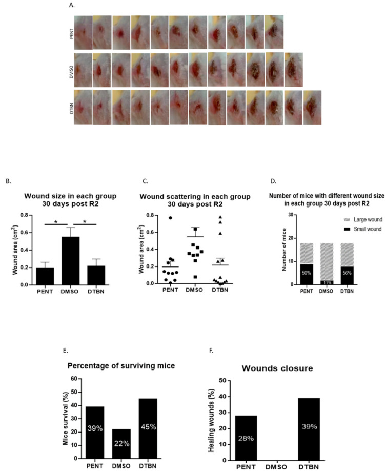
Cutaneous leishmaniasis (CL) is a zoonotic disease, manifested as chronic ulcers, potentially leaving unattractive scars. There is no preventive vaccination or optimal medication against leishmaniasis. Chemotherapy generally depends upon a small group of compounds, each with its own efficacy, toxicity, and rate of drug resistance. To date, no standardized, simple, safe, and highly effective regimen for treating CL exists. Therefore, there is an urgent need to develop new optimal medication for this disease. Sesquiterpen thio-alkaloids constitute a group of plant secondary metabolites that bear great potential for medicinal uses. The nupharidines found in Nuphar lutea belong to this group of compounds. We have previously published that Nuphar lutea semi-purified extract containing major components of nupharidines has strong anti-leishmanial activity in vitro. Here, we present in vivo data on the therapeutic benefit of the extract against Leishmania major (L. major) in infected mice. We also expanded these observations by establishing the therapeutic effect of the extract-purified nupharidine 6,6'-dihydroxythiobinupharidine (DTBN) in vitro against promastigotes and intracellular amastigotes as well as in vivo in L. major-infected mice. The results suggest that this novel anti-parasitic small molecule has the potential to be further developed against Leishmania.

Keywords: 6,6′-dihydroxythiobinupharidine (DTBN); Leishmania major; Nuphar lutea; anti-Leishmania small molecule; cutaneous leishmaniasis.


Conflict of interest statement

The authors declare no conflict-of-interest representation or interpretation of the reported research results.

Figures

Figure 1
Figure 1
Structure of 6,6′-dihydroxythiobinupharidine (DTBN).
Figure 2
Figure 2
Survival of DTBN-treated Lm promastigotes. First, 1 × 10⁶/well Leishmania major promastigotes in 1 mL were added to 96-well plates in a 200 µL final volume (200,000 parasites/well). Each well was treated with paromomycin (Paro.) as the gold standard, DTBN 0.1 µg/mL or DTBN 0.2 µg/mL or vehicle. Then, 48 h later, the motile (live) promastigotes were counted out of 100 parasites and the percentage of viable parasites was confirmed on a hemocytometer with trypan blue. The surviving promastigotes are presented as a percentage of untreated live promastigotes. Mean and SD were calculated.
Figure 3
Figure 3
Transmission (TEM) and scanning electron microscopy (SEM) images of L. major promastigotes treated with DTBN. L. major promastigotes were treated with growth media or 0.1 µg/mL DTBN for 24 h. The cells were then washed with PBS and processed as described for EM evaluation. (A1) Control, untreated promastigotes with SEM pictures (bar 5 mm), (A2) Untreated promastigotes, with TEM pictures. (B1) SEM pictures of treated promastigotes at two magnifications (left B1) (5.55K (bar 15 mm) and (right B1) 7.5K (bar 7.5 mm) magnification (bar 7.5 mm)). Arrows show damaged promastigotes, some lack flagella or show cell body and organelle damage. (B2) TEM of promastigotes treated with 0.1 µg/mL DTBN. M—mitochondria; N—nucleus; F—flagella; K—kinetoplast.
Figure 4
Figure 4
DTBN treatment of infected macrophages reduces the amount of intracellular L. major amastigotes. First, 24-well plates were covered with slides. Then, 3 × 10⁵/well C3H peritoneal macrophages were left to adhere for 3 h on glass-slide-covered wells. Next, 1 × 10⁶/well L. major promastigotes were added to each well (in triplicate). After 24 h, cells were treated (A) vehicle, (B) with paromomycin (Paro) 200 µg/mL, (C) DTBN 2 µg/mL or (D) DTBN 5 µg/mL. After 72 h, the slides were Giemsa stained and the intracellular amastigotes are shown by the arrow. Pictures were taken at a ×1000 magnification. (E) In parallel, 72 h later, equally treated macrophage cultures were collected, and cDNA was synthesized. The DNA samples were run in multiplex hydrolysis probe-based real-time PCR (mqRT-PCR). L. major cDNA relative amounts as compared to the positive control paromomycine were calculated based on a promastigote DNA calibration curve. The results were normalized to vehicle-treated values. The Leishmania-infected macrophage cDNA sample value was 1. Thus, 2 µg/mL was not effective and 3 and 5 µg/mL showed a dose response of about 10% and 70% effectiveness.
Figure 5
Figure 5
Nuphar lutea semi-purified extract (NUP.E) reduces the size of Lm wounds in treated mice. First, 1 × 10⁸/100 µL L. major promastigotes were injected into the tail base of 40 Balb/c male mice. Ten days later, following the appearance of the wound, 20 mg/kg/mouse NUP.E or acidic water (vehicle) was IP injected daily into mice for 8 days. Small wounds were defined as <0.1 cm2; large wounds as >0.1 cm2. The wound size was determined by the Digimizer program. Pictures of the wounds were taken at days 0 and 8 after treatment as well as 5 and 30 days post-treatment. (A) Representative pictures of differences in wound size between treated and control groups. (B) Proportion of mice with small/large wound area after 8 days of treatment. (C) Proportion of mice with small/large wound area 5 days post-treatment. (D) Wound area scattering of treated mice (each dot represents the wound area of one mouse in the experiment) as compared to control, 5 days post-treatment (t-test; p < 0.0016, ** p < 0.01). (E) Wound area scattering of treated mice as compared to control, 30 days post-treatment. No significant difference in wound size was observed between the two groups. Mice were sacrificed when the wound area reached 1.5 cm2.
Figure 6
Figure 6
Reduction in in vivo wound size of L. major-infected mice by intra-lesion (IL) injection of DTBN or vehicle. First, 1 × 10⁸/100 µL L. major promastigotes were injected into the mouse tail base. Next, 10 days afterward, the lesions appeared and 20 µg/40 µL of DTBN or vehicle (diluted DMSO) (n = 18) was IL injected. As a positive control, 20 mg/kg/mouse Pentostam (PENT) (n = 18) was IP injected. All treatments were given once a day for 15 days (R1) and the treatment was then stopped for 12 days and resumed for 7 more days (R2). Wound pictures were taken 30 days post-R2. (A) Increasing wound size from the smallest to the largest in each group, 30 days post-R2. (B) Wound size 30 days post-R2 of mice treated with DTBN or Pentostam as compared to the vehicle group. (Mean and SEM were calculated by Anova test; p < 0.0128, * p < 0.05). (C) Wound size distribution 30 days post-R2. (D) Proportion of mice with large/small wounds 30 days post-R2. The number of mice in each group was 18 (small wound: cm2 < 0.1; big wound: cm2 > 0.1 The wound size was analyzed by the Digimizer program). (E) The proportion of mice who survived in each group (18 mice/group) was determined at the endpoint of the experiment, 48 days post-R2. Mice were sacrificed when the wound area reached 1.5 cm2. (F) Wound size was compared at 48 days post-R2 to the wound size at 30 days post-R2. Unchanged wound size or size reduction was considered a cured wound.

References

    1. Corman H.N., McNamara C.W., Bakowski M.A. Drug Discovery for Cutaneous Leishmaniasis: A Review of Developments in the Past 15 Years. Microorganisms. 2023;11:2845. doi: 10.3390/MICROORGANISMS11122845. - DOI - PMC - PubMed
    1. De Vries H.J.C., Schallig H.D., De Vries H.J.C., Schallig H.D. Cutaneous Leishmaniasis: A 2022 Updated Narrative Review into Diagnosis and Management Developments. Am. J. Clin. Dermatol. 2022;23:823–840. doi: 10.1007/s40257-022-00726-8. - DOI - PMC - PubMed
    1. Leishmaniasis. [(accessed on 12 February 2024)]. Available online: https://www.who.int/health-topics/leishmaniasis#tab=tab_1.
    1. Dayakar A., Chandrasekaran S., Kuchipudi S.V., Kalangi S.K. Cytokines: Key Determinants of Resistance or Disease Progression in Visceral Leishmaniasis: Opportunities for Novel Diagnostics and Immunotherapy. Front. Immunol. 2019;1:670. doi: 10.3389/fimmu.2019.00670. - DOI - PMC - PubMed
    1. Porta E.O., Gao L., Denny P.W., Steel P.G., Kalesh K. Inhibition of HSP90 distinctively modulates the global phosphoproteome of Leishmania mexicana developmental stages. Microbiol. Spectr. 2023;11:e0296023. doi: 10.1128/spectrum.02960-23. - DOI - PMC - PubMed

MeSH terms

LinkOut - more resources