. 2024 May 14;13(5):405.
doi: 10.3390/pathogens13050405.

Role of Type 4B Secretion System Protein, IcmE, in the Pathogenesis of Coxiella burnetii

Affiliations

Role of Type 4B Secretion System Protein, IcmE, in the Pathogenesis of Coxiella burnetii

Rajesh Palanisamy et al. Pathogens. .

Abstract

Coxiella burnetii is an obligate intracellular Gram-negative bacterium that causes Q fever, a life-threatening zoonotic disease. C. burnetii replicates within an acidified parasitophorous vacuole derived from the host lysosome. The ability of C. burnetii to replicate and achieve successful intracellular life in the cell cytosol is vastly dependent on the Dot/Icm type 4B secretion system (T4SSB). Although several T4SSB effector proteins have been shown to be important for C. burnetii virulence and intracellular replication, the role of the icmE protein in the host-C. burnetii interaction has not been investigated. In this study, we generated a C. burnetii Nine Mile Phase II (NMII) mutant library and identified 146 transposon mutants with a single transposon insertion. Transposon mutagenesis screening revealed that disruption of icmE gene resulted in the attenuation of C. burnetii NMII virulence in SCID mice. ELISA analysis indicated that the levels of pro-inflammatory cytokines, including interleukin-1β, IFN-γ, TNF-α, and IL-12p70, in serum from Tn::icmE mutant-infected SCID mice were significantly lower than those in serum from wild-type (WT) NMII-infected mice. Additionally, Tn::icmE mutant bacteria were unable to replicate in mouse bone marrow-derived macrophages (MBMDM) and human macrophage-like cells (THP-1). Immunoblotting results showed that the Tn::icmE mutant failed to activate inflammasome components such as IL-1β, caspase 1, and gasdermin-D in THP-1 macrophages. Collectively, these results suggest that the icmE protein may play a vital role in C. burnetii virulence, intracellular replication, and activation of inflammasome mediators during NMII infection.

Keywords: Coxiella burnetii; caspase 1; cytokines; icmE protein; inflammasome; pyroptosis; type IV secretion system; virulence.


Conflict of interest statement

The authors declare no conflicts of interest.

Figures

Figure 1
Figure 1
Construction of a C. burnetii NMII RSA439 transposon mutant library. (A) The pITR-CAT-ColE1-P311 transposable plasmid was employed to generate a Coxiella mutant library containing the tetracycline-resistant gene under the regulation of the Coxiella promoter p1169, flanked by Inverted Terminal Repeats (ITR). The blue curved arrow indicates the site of the nucleotide sequence encoding a lysine for autotropic-based selection. (B) Schematic presentation of the total number of transposon mutants obtained, trimmed, and annotated in the Coxiella NMII RSA439 genome. (C) The bar graph represents the location of each transposon insertion in the genome, and the bar height shows the number of mutants with a transposon insertion at each site.
Figure 2
Figure 2
Bioinformatics analysis of C. burnetii icmE nucleotide and protein sequences. (A) Location of transposon insertions that disrupted icmE in the C. burnetii NMII RSA439 genome. The C. burnetii icmE gene is represented in the red frame, and the deduced protein and its domain profile are shown in the black solid frame. (B) Multiple sequence alignment of the TrbI domain of C. burnetii icmE with its orthologs. Strongly conserved sequences are highlighted by * symbols. (C) The predicted 3D structure of the C. burnetii icmE protein was viewed using PyMOL. The TrbI domain was highlighted in a dotted circle, and conserved motifs such as ‘NSD’ (purple) and ‘ART’ (sea blue) are shown in the ball model.
Figure 3
Figure 3
C. burnetii icmE is required for intracellular replication in MBMDM and THP-1 cells. The transposon icmE mutant was evaluated for its intracellular replication ability in MBMDM. MBMDM were infected with 100 MOI of WT NMII, Tn::dot, Tn::icmE or Tn::icmE complement in poly D-lysine-coated glass slide chambers. Following the infection for 3 days, all cells were fixed and stained with antibodies against Coxiella [46], LAMP1 (green) and Hoechst dye (blue) and examined by confocal fluorescence microscopy at magnification of 40×. (A) Confocal images of bacterial intracellular replication of WT NMII, Tn::dotA, Tn::icmE and Tn::icmE complement strains in MBMDM. C. burnetii bacterial numbers in MBMDM (B) and THP-1 macrophages (D) were determined by real-time qPCR and are expressed as C. burnetii com1 gene copy numbers. In terms of com1 gene copy number in genomic equivalent at 3 dpi. Percentage of vacuole formation in MBMDM (C) and THP-1 macrophages (E) at 3 dpi. Error bars indicate the mean ± standard deviation, and the results are expressed as the mean of three individual experiments, conducted with biological duplicates and three technical replicates. The p-value was calculated using an unpaired t-test. p < 0.01 **; p < 0.001 ***; and p < 0.0001 ****.
Figure 4
Figure 4
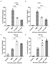
Compare WT NMII-, Tn::dotA- and Tn::icmE-infection-induced cytokine responses in MBMDM. MAGPIX Luminex analysis of IL-1β and TGF-β1 cytokines in culture supernatants from MBMDM infected with WT NMII, Tn::dotA, or Tn::icmE at MOI of 100 at 24 and 48 hpi. Compared to WT NMII-infected cells, Tn::icmE or Tn::dotA-infected cells had lower levels of IL-1β secretion (A,B) at 24 and 48 hpi, and increased the level of TGF-β1 secretion (C,D) at 24 and 48 hpi. Error bars indicate the mean ± standard deviation, and the results are expressed as the mean of three individual experiments, conducted with biological duplicates and three technical replicates. The p-value was calculated using an unpaired t-test. p < 0.05 *; p < 0.01 **; and p < 0.001 ***.
Figure 5
Figure 5
C. burnetii icmE protein is required for activation of caspase 1-dependent inflammasome in THP-1 macrophages. THP-1-derived macrophages were infected with uninfected or infected with WT NMII, Tn:icmE or Tn::dotA at an MOI of 100. (A) Immunoblot of the inflammasome pathway-related proteins, caspase 1, cleaved caspase 1, IL-1β, Gasdermin and cleaved Gasdermin in the supernatants from uninfected, WT NMII-, Tn::icmE- or Tn::dotA-infected THP-1 macrophages at 48 hpi. Relative intensity unites of caspase 1 (B), cleaved caspase 1 (C), IL-1β (D), Gasdermin (E) and cleaved Gasdermin (F). The p-value was calculated using an unpaired t-test. p < 0.05 *; and p < 0.005 ***. The symbols p < 0.05, *, and p < 0.001, *** are used to denote the presence of significant differences across samples, as determined by the unpaired t-test. The error bars represent the standard deviations of the means. The experiment was performed in triplicate.
Figure 6
Figure 6
Evaluate the severity of Tn::icmE mutant infection-induced disease in SCID mice. The SCID mice were infected with 1 × 10⁹ GE of WT NMII, Tn::dotA, Tn::icmE or Tn::icmE complement. (A) Relative body weights (current body weight/day 0 body weight) were measured throughout the challenge period. (B) Splenomegaly (% of spleen weight/body weight). (C) bacterial burden in the spleen was determined by real-time qPCR and is expressed as C. burnetii com1 gene copy numbers. Each experimental group consists of four mice, and the error bars indicate the standard deviations from the means. The symbol p < 0.05, *; p < 0.01, **; p < 0.001, *** was used to denote the presence of significant differences across samples, as determined by the unpaired t-test.
Figure 7
Figure 7
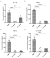
The Tn::icmE mutant induced lower levels of pro-inflammatory cytokine responses in SCID mice. The SCID mice were infected intraperitoneally with 109 GE of WT NMII, Tn::dotA or Tn::icmE. Serum cytokine concentrations of IL-1β (A), IFN-γ (B), TNF-α (C), and IL-12p70 (D) were measured using the MAGPIX Luminex xMAP instrument at 14 dpi. Each experimental group consists of four mice, and the error bars indicate the standard deviations from the means. The symbols p < 0.01,**, p < 0.001,***, and p < 0.0001,**** are used to denote the presence of significant differences across samples, as determined by the unpaired t-test.

Similar articles

Cited by

  • Navigating Q fever: Current perspectives and challenges in outbreak preparedness.
    Meles DK, Khairullah AR, Mustofa I, Wurlina W, Akintunde AO, Suwasanti N, Mustofa RI, Putra SW, Moses IB, Kusala MKJ, Raissa R, Fauzia KA, Aryaloka S, Fauziah I, Yanestria SM, Wibowo S. Meles DK, et al. Open Vet J. 2024 Oct;14(10):2509-2524. doi: 10.5455/OVJ.2024.v14.i10.2. Epub 2024 Oct 31. Open Vet J. 2024. PMID: 39545195 Free PMC article. Review.

References

    1. Van Schaik E.J., Case E.D., Martinez E., Bonazzi M., Samuel J.E. The SCID Mouse Model for Identifying Virulence Determinants in Coxiella burnetii. Front. Cell Infect. Microbiol. 2017;7:25. doi: 10.3389/fcimb.2017.00025. - DOI - PMC - PubMed
    1. Maurin M., Raoult D. Q Fever. Clin. Microbiol. Rev. 1999;12:518–553. doi: 10.1128/cmr.12.4.518. - DOI - PMC - PubMed
    1. Raoult D., Marrie T.J., Mege J.L. Natural history and pathophysiology of Q fever. Lancet Infect. Dis. 2005;5:219–226. doi: 10.1016/S1473-3099(05)70052-9. - DOI - PubMed
    1. Parker N.R., Barralet J.H., Bell A.M. Q fever. Lancet. 2006;367:679–688. doi: 10.1016/S0140-6736(06)68266-4. - DOI - PubMed
    1. CDC . Q Fever. Centers for Disease Control and Prevention; Antlanta, GA, USA: 2019. [(accessed on 25 June 2023)]. Available online: https://www.cdc.gov/qfever/

MeSH terms

LinkOut - more resources