. 2024 May 15;13(5):410.
doi: 10.3390/pathogens13050410.

Leucoverdazyls as Novel Potent Inhibitors of Enterovirus Replication

Affiliations

Leucoverdazyls as Novel Potent Inhibitors of Enterovirus Replication

Alexandrina S Volobueva et al. Pathogens. .

Abstract

Enteroviruses (EV) are important pathogens causing human disease with various clinical manifestations. To date, treatment of enteroviral infections is mainly supportive since no vaccination or antiviral drugs are approved for their prevention or treatment. Here, we describe the antiviral properties and mechanisms of action of leucoverdazyls-novel heterocyclic compounds with antioxidant potential. The lead compound, 1a, demonstrated low cytotoxicity along with high antioxidant and virus-inhibiting activity. A viral strain resistant to 1a was selected, and the development of resistance was shown to be accompanied by mutation of virus-specific non-structural protein 2C. This resistant virus had lower fitness when grown in cell culture. Taken together, our results demonstrate high antiviral potential of leucoverdazyls as novel inhibitors of enterovirus replication and support previous evidence of an important role of 2C proteins in EV replication.

Keywords: 2C protein; antioxidant; antiviral; coxsackievirus; enteroviruses; leucoverdazyls.


Conflict of interest statement

The authors declare no conflicts of interest.

Figures

Figure 1
Figure 1
Structures of leucoverdazyls tested in the study and reference compounds: pleconaril and guanidine hydrochloride. Pleconaril was kindly provided by Dr. V. A. Makarov (Research Center of Biotechnology RAS, 33-1 Leninsky Prospect, 119071, Moscow, Russia). Guanidine hydrochloride was bought from Dia-M Ltd. (Moscow, Russia).
Figure 2
Figure 2
Antioxidant activity of selected dihydrotetrazines. Presented are IC50 values (µM) for the antioxidant activity of the tested compounds by DPPH assay. Vitamin C was used as a reference.
Figure 3
Figure 3
Thermostabilizing properties of 1a in comparison to those of pleconaril. Values are the mean ± SD of three independent experiments. The legend shows the concentration of each compound tested. The asterisk indicates the significance of the difference in viral titer for pleconaril at 51 °C and 55.2 °C relative to the virus control, p < 0.05 by Mann–Whitney U-test.
Figure 4
Figure 4
Results of time-of-addition assay for 1a. The activity of compound 1a against the Coxsackie B4 virus (Powers strain) depending on the time of addition to a permissive cell line upon CVB4 infection. Vero cells were infected with CVB4 (−1 h), and 1a (5 μg/mL) was added at the indicated time points (in hours) either before the virus (−2 h), concomitantly with the virus (−1 h), or after (0, 2, 4, 6 h) infection, where 0 corresponds to the moment of completed virus absorption on the cell surface. The infectious activity of the viral progeny was evaluated by end-point titration in the Vero cells in lg TCID50/0.2 mL. Pleconaril (10 μg/mL) was used as a reference compound. Values are presented as the mean ± SD of three independent experiments. An asterisk indicates a significant difference in viral titer for 1a and pleconaril relative to the virus control, p < 0.05 by the Mann–Whitney U-test.
Figure 5
Figure 5
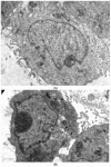
Ultrastructure of Vero cells infected by CVB3 revealed by transmissive electron microscopy, representative microphotographs. (A) Intact cell. No vacuoles or replication organelles are visible within the cytoplasm. (B) CVB3-infected cell. Numerous vacuoles representing virus-specific replication organelles are indicated by arrowheads. (C) CVB3-infected cell in the presence of 100 μM compound 1a. No morphological signs of viral replication can be seen. (D) Statistical analysis of cell numbers with and without signs of viral replication in 1a treated CVB3 infected group versus CVB3 infected non-treated group, df = 1, N = 110, χ2 = 39.79, p < 0.05.
Figure 5
Figure 5
Ultrastructure of Vero cells infected by CVB3 revealed by transmissive electron microscopy, representative microphotographs. (A) Intact cell. No vacuoles or replication organelles are visible within the cytoplasm. (B) CVB3-infected cell. Numerous vacuoles representing virus-specific replication organelles are indicated by arrowheads. (C) CVB3-infected cell in the presence of 100 μM compound 1a. No morphological signs of viral replication can be seen. (D) Statistical analysis of cell numbers with and without signs of viral replication in 1a treated CVB3 infected group versus CVB3 infected non-treated group, df = 1, N = 110, χ2 = 39.79, p < 0.05.
Figure 6
Figure 6
Comparison of IC50 values for the CVB3 R and CVB3 WT strains. Presented are the results of the viral yield reduction assay for two CVB3 strains: wild-type and resistant virus propagated in the presence of 1a. The 4PL were fitted using GraphPad Prism 6. Viral titer is represented in % relative to virus control.
Figure 7
Figure 7
Propagation kinetics of CVB3 WT and CVB3 R strains in Vero cells with and without 1a. Viral progeny titer is plotted versus incubation time. Multistep growth curves are presented. An asterisk indicates a significant difference in virus titer, p < 0.05 by the Mann–Whitney U-test.
Figure 8
Figure 8
Position of S109I mutation in 2C protein. The amino acid position is depicted in red. The C- and N- termini of the protein are marked with C and N, respectively.
Figure 9
Figure 9
Colocalization of the 1a binding site and the S109I amino acid substitution in the 2C protein of 1a-resistant Coxsackievirus B3. Different protein chains are marked with different colors. The substitution is indicated by the arrow.

References

    1. Simmonds P., Gorbalenya A.E., Harvala H., Hovi T., Knowles N.J., Lindberg A.M., Oberste M.S., Palmenberg A.C., Reuter G., Skern T., et al. Recommendations for the nomenclature of enteroviruses and rhinoviruses. Arch. Virol. 2020;165:793–797. doi: 10.1007/s00705-019-04520-6. Erratum in Arch. Virol. 2020, 165, 1515. - DOI - PMC - PubMed
    1. Lugo D., Krogstad P. Enteroviruses in the early 21st century: New manifestations and challenges. Curr. Opin. Pediatr. 2016;28:107–113. doi: 10.1097/MOP.0000000000000303. - DOI - PMC - PubMed
    1. Genoni A., Canducci F., Rossi A., Broccolo F., Chumakov K., Bono G., Salerno-Uriarte J., Salvatoni A., Pugliese A., Toniolo A. Revealing enterovirus infection in chronic human disorders: An integrated diagnostic approach. Sci. Rep. 2017;7:5013. doi: 10.1038/s41598-017-04993-y. - DOI - PMC - PubMed
    1. Nekoua M.P., Alidjinou E.K., Hober D. Persistent coxsackievirus B infection and pathogenesis of type 1 diabetes mellitus. Nat. Rev. Endocrinol. 2022;18:503–516. doi: 10.1038/s41574-022-00688-1. - DOI - PMC - PubMed
    1. Chapman N.M. Persistent Enterovirus Infection: Little Deletions, Long Infections. Vaccines. 2022;10:770. doi: 10.3390/vaccines10050770. - DOI - PMC - PubMed

MeSH terms

LinkOut - more resources