. 2024 May 24;20(5):e1012266.
doi: 10.1371/journal.ppat.1012266. eCollection 2024 May.

Mycoplasma glycine cleavage system key subunit GcvH is an apoptosis inhibitor targeting host endoplasmic reticulum

Affiliations

Mycoplasma glycine cleavage system key subunit GcvH is an apoptosis inhibitor targeting host endoplasmic reticulum

Qiao Pan et al. PLoS Pathog. .

Abstract

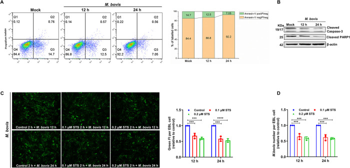
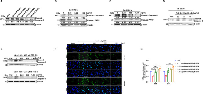
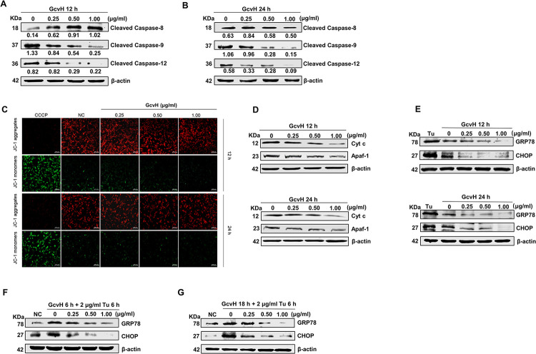
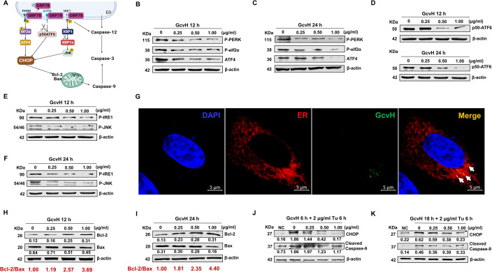
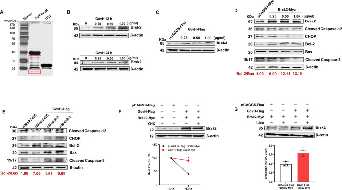
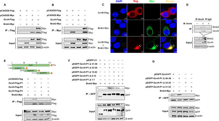
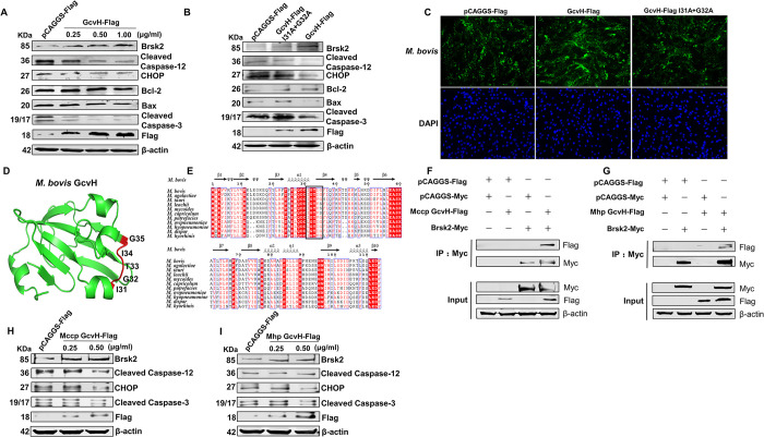
Mycoplasmas are minimal but notorious bacteria that infect humans and animals. These genome-reduced organisms have evolved strategies to overcome host apoptotic defense and establish persistent infection. Here, using Mycoplasma bovis as a model, we demonstrate that mycoplasma glycine cleavage system (GCS) H protein (GcvH) targets the endoplasmic reticulum (ER) to hijack host apoptosis facilitating bacterial infection. Mechanically, GcvH interacts with the ER-resident kinase Brsk2 and stabilizes it by blocking its autophagic degradation. Brsk2 subsequently disturbs unfolded protein response (UPR) signaling, thereby inhibiting the key apoptotic molecule CHOP expression and ER-mediated intrinsic apoptotic pathway. CHOP mediates a cross-talk between ER- and mitochondria-mediated intrinsic apoptosis. The GcvH N-terminal amino acid 31-35 region is necessary for GcvH interaction with Brsk2, as well as for GcvH to exert anti-apoptotic and potentially pro-infective functions. Notably, targeting Brsk2 to dampen apoptosis may be a conserved strategy for GCS-containing mycoplasmas. Our study reveals a novel role for the conserved metabolic route protein GcvH in Mycoplasma species. It also sheds light on how genome-reduced bacteria exploit a limited number of genomic proteins to resist host cell apoptosis thereby facilitating pathogenesis.


Conflict of interest statement

The authors have declared that no competing interests exist.

Figures

Fig 1
Fig 1. Anti-apoptotic effect of M. bovis.
(A) Apoptosis analysis in M. bovis-infected cells by flow cytometry with dual Annexin V-PI cell labeling. Q4 quadrants represent intact cells (Annexin V negative\PI negative); Q3 quadrants represent early apoptotic cells (Annexin V positive\PI negative); Q2 quadrants indicate late apoptotic cells (Annexin V positive\PI positive); and Q1 quadrants indicate necrotic cells (Annexin V negative\PI positive). The graph on the right represents the percentage of apoptotic and intact cells, and the non-significant percentages of Annexin V-negative and PI-positive cells were excluded. (B) The cleaved levels of Caspase-3 and PARP1 were downregulated in M. bovis-infected EBL cells. (C-D) The percentage of cell-associated M. bovis significantly decreased in EBL cells pretreated with STS. All assays were performed with three independent experiments, and values represent the means ± SDs. Significance was assessed by one-way ANOVA with Dunnett’s multiple comparison tests relative to the control. ***, p < 0.001; ****, p < 0.0001.
Fig 2
Fig 2. M. bovis encodes an anti-apoptotic protein GcvH.
(A) The group-D MAPs decreased cleaved Caspase-3 levels by Western blotting analysis. (B-C) Western blotting revealed that the cleaved levels of Caspase-3 and PARP1 were reduced following GcvH protein incubation in EBL cells. (D) GcvH blockage increased M. boivs apoptotic effect on EBL cells. M. bovis was incubated with the purified antibodies against GcvH (10, 20 and 50 μg/ml) or PBS for 30 min and then added to EBL cells. The cleaved Caspase-3 level in EBL cells was determined by Western blotting. (E-F) GcvH lowered or prevented STS-induced apoptosis in EBL cells. EBL cells were treated with 0, 0.25, 0.5 or 1.00 μg/ml GcvH for 6 or 18 h, and then with 0.25 μM STS. Cells were collected for Western blotting analysis of cleaved Caspase-3 level (E), or for TUNEL detection (F). (G) EBL cells were treated as described in panel E and then incubated with M. bovis for 12 h. The number of M. bovis per EBL cells was surveyed by TaqMan qPCR. The protein levels were quantified by ImageJ and normalized to β-actin (A, B and C). The data are presented as the means ± SDs from three independent experiments, and significance was assessed by one-way ANOVA with Tukey’s multiple comparison test. **, p < 0.01; ***, p < 0.001; ****, p < 0.0001.
Fig 3
Fig 3. GcvH resists host cell apoptosis through intrinsic apoptotic signaling.
(A-B) EBL cells were mocked or incubated with GcvH for 12 or 24 h. Cell lysates were analyzed by western blotting for cleaved Caspase-8, -9, -12 and β-actin. The protein levels were quantified by ImageJ and normalized to β-actin. (C) The mitochondrial membrane potential (MMP) of GcvH-incubated EBL cells was measured by the JC-1 probe, CCCP as positive control. (D) The levels of Cyt c and Apaf-1 in GcvH-incubated EBL cells were evaluated by Western blotting. (E) GcvH treatment decreased the levels of GRP78 and CHOP. (F-G) Tunicamycin (Tu)-induced increases in GRP78 and CHOP expression were reduced or even abolished by GcvH.
Fig 4
Fig 4. GcvH disturbs three UPR branches to block ER-mediated apoptosis.
(A) Schematic showing CHOP activated by the three UPR branches in the ER. (B-C) The inhibition of the PERK branch in EBL cells following GcvH incubation was confirmed by Western blotting of the levels of phosphorylation-PERK, -eIF2α and ATF4. (D) The downregulation of ATF6 branch in EBL cells with GcvH addition was determined by Western blotting of ATF6 cleavage (p50 ATF6). (E-F) The suppression of the IRE1 pathway in EBL cells following GcvH incubation was obtained by Western blotting of the phosphorylation level of IRE1 and JNK. (G) EBL cells were incubated with 1 μg/ml GcvH protein for 12 h, and then intracellular GcvH protein (green) was localized using the purified specific anti-GcvH antibody conjugated with an anti-mouse fluorescent secondary antibody. The EBL cell’s endoplasmic reticulum (ER, red) was labeled by transfecting with the plasmid pDsRed2-ER. DAPI was used to stain cellular nuclei (blue). (H-I) GcvH-induced enhancement of the Bcl-2/Bax ratio (red characters beneath the WB bands) was verified by Western blotting for the level of Bcl-2 and Bax. (J-K) EBL cells were incubated with GcvH (0, 0.25, 0.5 and 1 μg/ml) for 6 or 18 h, and followed by an addition of 2 μg/ml Tu for an additional 6 h. Cell lysates were tested by western blotting for CHOP, cleaved Caspase-9 and β-actin. The protein levels were quantified by ImageJ and normalized to β-actin (H, I, J and K).
Fig 5
Fig 5. BRSK2 is essential for the anti-apoptotic effect of GcvH.
(A) Screening for the host proteins interacting with GcvH by the GST pull-down assay. (B-C) GcvH-induced promotion of Brsk2 expression was confirmed by Western blotting of the Brsk2 and β-actin levels in EBL cells with GcvH protein incubation (B) or with transfection of the GcvH-Flag plasmid (C). (D) The inhibition of ER-associated intrinsic apoptotic pathway and apoptosis in EBL cells transfected with the GcvH-Flag plasmid were ascertained by Western blotting of the levels of cleaved Caspase-12, -3, CHOP, Bcl-2, Bax, and β-actin. (E) Knockdown of Brsk2 disrupted the inhibitory effect of GcvH on EBL cell apoptosis. (F) GcvH stabilized Brsk2 by preventing its degradation. EBL cells coexpressing Brsk2-Myc and GcvH-Flag or empty vector control (Flag) were treated with the protein synthesis inhibitor cycloheximide (CHX; 75 μg/ml) for 12 h. Brsk2 and β-actin levels were determined by Western blotting analysis. Densitometry analysis of Brsk2 levels relative to actin (Brsk2/actin) normalized to no CHX treatment. (G) GcvH augmented Brsk2 stability by diminishing the autophagic degradation of Brsk2. EBL cells coexpressing Brsk2-Myc and GcvH-Flag or empty vector control (Flag) were treated with an autophagy inhibitor 3-MA (5 mM, 12 h). Brsk2 and β-actin levels were determined by Western blotting analysis. Densitometry analysis of Brsk2 levels relative to actin (Brsk2/actin) in 3-MA-treated or -untreated EBL cells. The ratio of 3-MA-treated (Brsk2/actin) was normalized to 3-MA-untreated (Brsk2/actin). The data represented the means ± SDs of the results from three independent experiments. Significance was assessed by a two-tailed Student’s t test. *, p < 0.05; **, p < 0.01.
Fig 6
Fig 6. GcvH N-terminal mutations (aa 31 to 35) abolish its interaction with Brsk2.
(A-B) GcvH-Flag coimmunoprecipitated with Brsk2-Myc. Hela cells were transfected with GcvH-Flag or Brsk2-Myc, or the control plasmids pCAGGS-Flag or pCAGGS-Myc for 36 h. Cell lysates were collected for Co-IP with beads conjugated with anti-Flag antibody (A) and Myc antibody (B). (C) Colocalization of the GcvH and Brsk2 proteins. (D) Hela cells were infected with M. bovis and the interaction of endogenous Brsk2 and GcvH protein in the cell lysates was detected by coimmunoprecipitation with antibody against GcvH proteins rather than IgG and protein A-conjugated beads. (E) The fragment of GcvH aa 1–38 interacted with the Brsk2 protein. Upper: Schematic of the GcvH truncates tagged with Flag and locations of the aa residues are noted. Lower: Hela cells were co-transfected with Brsk2-Myc and truncated GcvH constructs, and the cell lysates were collected for Co-IP assays using beads conjugated with anti-Flag antibody for Flag-tagged GcvH truncates or with Myc antibody for Myc-tagged Brsk2. (F) The GcvH plasmid lacking the aa 31 to 38 did not immunoprecipitate with the Brsk2. Hela cells were cotransfected with Brsk2-Myc and the five GcvH-P1 deletion constructs. Cell lysates were collected for Co-IP assays using beads conjugated with anti-GFP antibodies. (G) The GcvH plasmid lacking the aa 31 to 32 or 33 to 35 did not immunoprecipitate with the Brsk2. Hela cells were cotransfected with Brsk2-Myc and the three GcvH-P1 deletion constructs, and then collected to perform a Co-IP assay.
Fig 7
Fig 7. The interaction with Brsk2 is required for the anti-host apoptotic action of GcvH.
(A) Transfection of the GcvH-Flag plasmid in EBL cells enhanced Brsk2 expression while hindering cell apoptosis. (B) GcvH had no anti-apoptotic effects on host cells when it lost its binding to Brsk2. (C) GcvH did not promote M. bovis infection of EBL cells when it lost the interaction with Brsk2. EBL cells were treated as stated in panel B and then subjected to detect cell-associated M. bovis by an IFA. (D) The PDB structure of M. bovis GcvH was predicted by the Alphafold network. Full-length GcvH protein (green) and its key amino acid sites 31–35 interacting with Brsk2 are shown here in red. This structure was visualized in Pymol software. (E) A multiple sequence comparison of GcvH between GCS-containing mycoplasmas. (F-G) Both Mccp GcvH-Flag and Mhp GcvH-Flag coimmunoprecipitated with Brsk2-Myc. (H-I) Both Mccp GcvH-Flag and Mhp GcvH-Flag transfection decreased host cell apoptosis.
Fig 8
Fig 8. Model depicting the promotion of M. bovis epithelial infection by GcvH anti-host cell apoptosis.
M. bovis attaches to cells, followed by its membrane protein GcvH binding to the host Brsk2 to resist the ER-mediated apoptotic signaling, which is transmitted to mitochondria via CHOP. GcvH promotes M. bovis infection by preventing host cell apoptosis through these two intrinsic apoptotic pathways.

Similar articles

References

    1. Citti C, Blanchard A. Mycoplasmas and their host: emerging and re-emerging minimal pathogens. Trends in microbiology. 2013;21(4):196–203. doi: 10.1016/j.tim.2013.01.003 - DOI - PubMed
    1. Razin S, Yogev D, Naot Y. Molecular biology and pathogenicity of mycoplasmas. Microbiology and molecular biology reviews. 1998;62(4):1094–156. doi: 10.1128/MMBR.62.4.1094-1156.1998 - DOI - PMC - PubMed
    1. Maes D, Sibila M, Kuhnert P, Segalés J, Haesebrouck F, Pieters M. Update on Mycoplasma hyopneumoniae infections in pigs: knowledge gaps for improved disease control. Transboundary and emerging diseases. 2018;65:110–24. - PubMed
    1. Calcutt M, Lysnyansky I, Sachse K, Fox L, Nicholas R, Ayling R. Gap analysis of Mycoplasma bovis disease, diagnosis and control: An aid to identify future development requirements. Transboundary and emerging diseases. 2018;65:91–109. - PubMed
    1. Dawood A, Algharib SA, Zhao G, Zhu T, Qi M, Delai K, et al.. Mycoplasmas as host pantropic and specific pathogens: clinical implications, gene transfer, virulence factors, and future perspectives. Frontiers in cellular and infection microbiology. 2022;12:855731. doi: 10.3389/fcimb.2022.855731 - DOI - PMC - PubMed

MeSH terms