

Automated Prediction of Proximal Middle Cerebral Artery Occlusions in Noncontrast Brain Computed Tomography
- PMID: 38787932
- PMCID: PMC11122774
- DOI: 10.1161/STROKEAHA.123.045772
Automated Prediction of Proximal Middle Cerebral Artery Occlusions in Noncontrast Brain Computed Tomography
Abstract
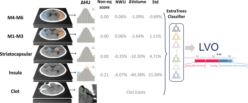
Background: Early identification of large vessel occlusion (LVO) in patients with ischemic stroke is crucial for timely interventions. We propose a machine learning-based algorithm (JLK-CTL) that uses handcrafted features from noncontrast computed tomography to predict LVO.
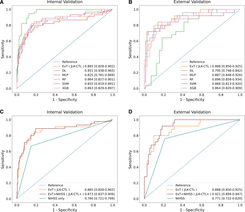
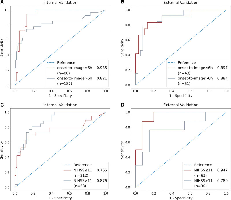
Methods: We included patients with ischemic stroke who underwent concurrent noncontrast computed tomography and computed tomography angiography in seven hospitals. Patients from 5 of these hospitals, admitted between May 2011 and March 2015, were randomly divided into training and internal validation (9:1 ratio). Those from the remaining 2 hospitals, admitted between March 2021 and September 2021, were designated for external validation. From each noncontrast computed tomography scan, we extracted differences in volume, tissue density, and Hounsfield unit distribution between bihemispheric regions (striatocapsular, insula, M1-M3, and M4-M6, modified from the Alberta Stroke Program Early Computed Tomography Score). A deep learning algorithm was used to incorporate clot signs as an additional feature. Machine learning models, including ExtraTrees, random forest, extreme gradient boosting, support vector machine, and multilayer perceptron, as well as a deep learning model, were trained and evaluated. Additionally, we assessed the models' performance after incorporating the National Institutes of Health Stroke Scale scores as an additional feature.
Results: Among 2919 patients, 83 were excluded. Across the training (n=2463), internal validation (n=275), and external validation (n=95) datasets, the mean ages were 68.5±12.4, 67.6±13.8, and 67.9±13.6 years, respectively. The proportions of men were 57%, 53%, and 59%, with LVO prevalences of 17.0%, 16.4%, and 26.3%, respectively. In the external validation, the ExtraTrees model achieved a robust area under the curve of 0.888 (95% CI, 0.850-0.925), with a sensitivity of 80.1% (95% CI, 72.0-88.1) and a specificity of 88.6% (95% CI, 84.7-92.5). Adding the National Institutes of Health Stroke Scale score to the ExtraTrees model increased sensitivity (from 80.1% to 92.1%) while maintaining specificity.
Conclusions: Our algorithm provides reliable predictions of LVO using noncontrast computed tomography. By enabling early LVO identification, our algorithm has the potential to expedite the stroke workflow.
Keywords: artificial intelligence; atrial fibrillation; computed tomography angiography; deep learning; ischemic stroke; predictive value of tests.
Conflict of interest statement
Figures
References
-
- Morsi RZ, Elfil M, Ghaith HS, Aladawi M, Elmashad A, Kothari S, Desai H, Prabhakaran S, Al-Mufti F, Kass-Hout T. Endovascular thrombectomy for large ischemic strokes: a living systematic review and meta-analysis of randomized trials. J Stroke. 2023;25:214–222. doi: 10.5853/jos.2023.00752 - PMC - PubMed
-
- Olive-Gadea M, Crespo C, Granes C, Hernandez-Perez M, Perez de la Ossa N, Laredo C, Urra X, Carlos Soler J, Soler A, Puyalto P, et al. . Deep learning based software to identify large vessel occlusion on noncontrast computed tomography. Stroke. 2020;51:3133–3137. doi: 10.1161/STROKEAHA.120.030326 - PubMed
-
- Weyland CS, Papanagiotou P, Schmitt N, Joly O, Bellot P, Mokli Y, Ringleb PA, Kastrup A, Mohlenbruch MA, Bendszus M, et al. . Hyperdense artery sign in patients with acute ischemic stroke-automated detection with artificial intelligence-driven software. Front Neurol. 2022;13:807145. doi: 10.3389/fneur.2022.807145 - PMC - PubMed
-
- Potter CA, Vagal AS, Goyal M, Nunez DB, Leslie-Mazwi TM, Lev MH. CT for treatment selection in acute ischemic stroke: a code stroke primer. Radiographics. 2019;39:1717–1738. doi: 10.1148/rg.2019190142 - PubMed
