

Neuroimaging to Facilitate Clinical Trials in Huntington's Disease: Current Opinion from the EHDN Imaging Working Group
- PMID: 38788082
- PMCID: PMC11307036
- DOI: 10.3233/JHD-240016
Neuroimaging to Facilitate Clinical Trials in Huntington's Disease: Current Opinion from the EHDN Imaging Working Group
Abstract
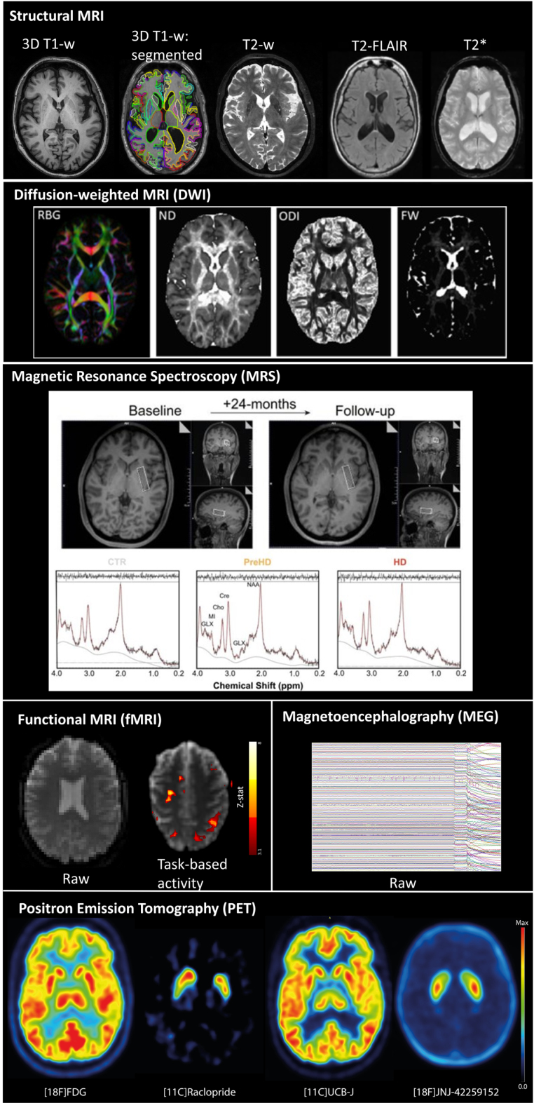
Neuroimaging is increasingly being included in clinical trials of Huntington's disease (HD) for a wide range of purposes from participant selection and safety monitoring, through to demonstration of disease modification. Selection of the appropriate modality and associated analysis tools requires careful consideration. On behalf of the EHDN Imaging Working Group, we present current opinion on the utility and future prospects for inclusion of neuroimaging in HD trials. Covering the key imaging modalities of structural-, functional- and diffusion- MRI, perfusion imaging, positron emission tomography, magnetic resonance spectroscopy, and magnetoencephalography, we address how neuroimaging can be used in HD trials to: 1) Aid patient selection, enrichment, stratification, and safety monitoring; 2) Demonstrate biodistribution, target engagement, and pharmacodynamics; 3) Provide evidence for disease modification; and 4) Understand brain re-organization following therapy. We also present the challenges of translating research methodology into clinical trial settings, including equipment requirements and cost, standardization of acquisition and analysis, patient burden and invasiveness, and interpretation of results. We conclude, that with appropriate consideration of modality, study design and analysis, imaging has huge potential to facilitate effective clinical trials in HD.
Keywords: Huntington’s disease; Neuroimaging; clinical trial; magnetic resonance imaging; magnetoencephalography; positron-emission tomography.
Conflict of interest statement
MP and KM are employees of IXICO plc. The authors have no other conflicts of interest to report.
Figures
Similar articles
-
Longitudinal neuroimaging biomarkers in Huntington's Disease.J Huntingtons Dis. 2013;2(1):21-39. doi: 10.3233/JHD-120030. J Huntingtons Dis. 2013. PMID: 25063427 Review.
-
Novel Imaging Biomarkers for Huntington's Disease and Other Hereditary Choreas.Curr Neurol Neurosci Rep. 2018 Oct 5;18(12):85. doi: 10.1007/s11910-018-0890-y. Curr Neurol Neurosci Rep. 2018. PMID: 30291526 Free PMC article. Review.
-
Huntington's disease: Brain imaging in Huntington's disease.Prog Mol Biol Transl Sci. 2019;165:321-369. doi: 10.1016/bs.pmbts.2019.04.004. Epub 2019 May 15. Prog Mol Biol Transl Sci. 2019. PMID: 31481169 Review.
-
Structural Magnetic Resonance Imaging in Huntington's Disease.Int Rev Neurobiol. 2018;142:335-380. doi: 10.1016/bs.irn.2018.09.006. Epub 2018 Oct 8. Int Rev Neurobiol. 2018. PMID: 30409258 Review.
-
Prognostic enrichment for early-stage Huntington's disease: An explainable machine learning approach for clinical trial.Neuroimage Clin. 2024;43:103650. doi: 10.1016/j.nicl.2024.103650. Epub 2024 Aug 10. Neuroimage Clin. 2024. PMID: 39142216 Free PMC article.
Cited by
-
Neuroimaging Techniques in Huntington's Disease: A Critical Review.Mov Disord Clin Pract. 2025 May;12(5):561-576. doi: 10.1002/mdc3.70010. Epub 2025 Feb 20. Mov Disord Clin Pract. 2025. PMID: 39976324 Review.
-
Advanced Magnetic Resonance Imaging for Early Diagnosis and Monitoring of Movement Disorders.Brain Sci. 2025 Jan 16;15(1):79. doi: 10.3390/brainsci15010079. Brain Sci. 2025. PMID: 39851446 Free PMC article. Review.
-
Microstructural Changes in the Corpus Callosum in Neurodegenerative Diseases.Cureus. 2024 Aug 21;16(8):e67378. doi: 10.7759/cureus.67378. eCollection 2024 Aug. Cureus. 2024. PMID: 39310519 Free PMC article. Review.
References
-
- A novel gene containing a trinucleotide repeat that is expanded and unstable on Huntington’s disease chromosomes. The Huntington’s Disease Collaborative Research Group. Cell. 1993;72(6):971–83. - PubMed
-
- Ghosh R, Tabrizi SJ. Clinical features of Huntington’s disease. Adv Exp Med Biol. 2018;1049:1–28. - PubMed
Publication types
MeSH terms
LinkOut - more resources
Full Text Sources
Medical
