. 2024 Jun 11;19(6):859-876.
doi: 10.1016/j.stemcr.2024.04.011. Epub 2024 May 23.

Human pluripotent stem cell-derived kidney organoids reveal tubular epithelial pathobiology of heterozygous HNF1B-associated dysplastic kidney malformations

Affiliations

Human pluripotent stem cell-derived kidney organoids reveal tubular epithelial pathobiology of heterozygous HNF1B-associated dysplastic kidney malformations

Ioannis Bantounas et al. Stem Cell Reports. .

Abstract

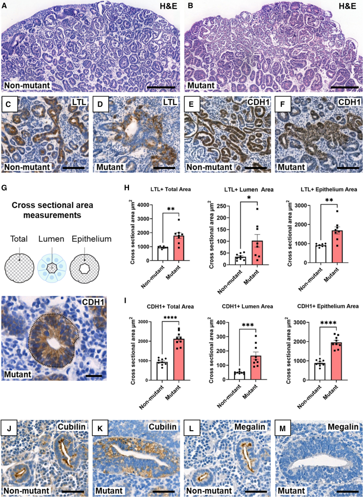
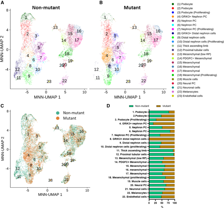
Hepatocyte nuclear factor 1B (HNF1B) encodes a transcription factor expressed in developing human kidney epithelia. Heterozygous HNF1B mutations are the commonest monogenic cause of dysplastic kidney malformations (DKMs). To understand their pathobiology, we generated heterozygous HNF1B mutant kidney organoids from CRISPR-Cas9 gene-edited human embryonic stem cells (ESCs) and induced pluripotent stem cells (iPSCs) reprogrammed from a family with HNF1B-associated DKMs. Mutant organoids contained enlarged malformed tubules displaying deregulated cell turnover. Numerous genes implicated in Mendelian kidney tubulopathies were downregulated, and mutant tubules resisted the cyclic AMP (cAMP)-mediated dilatation seen in controls. Bulk and single-cell RNA sequencing (scRNA-seq) analyses indicated abnormal Wingless/Integrated (WNT), calcium, and glutamatergic pathways, the latter hitherto unstudied in developing kidneys. Glutamate ionotropic receptor kainate type subunit 3 (GRIK3) was upregulated in malformed mutant nephron tubules and prominent in HNF1B mutant fetal human dysplastic kidney epithelia. These results reveal morphological, molecular, and physiological roles for HNF1B in human kidney tubule differentiation and morphogenesis illuminating the developmental origin of mutant-HNF1B-causing kidney disease.

Keywords: CRISPR; GRIK3; HNF1B; cAMP; glutamate receptors; kidney RNA-seq; kidney disease; organoid; pluripotent stem cells; proximal tubule.


Conflict of interest statement

Declaration of interests The authors declare no conflicts of interest.

Figures

None
Graphical abstract
Figure 1
Figure 1
HNF1B in hESC-derived organoids (A) RT-qPCR using HNF1B exon 2 primers showed similar upregulation in mutant and isogenic non-mutant lines over 25 days; bars denote standard error of the mean (SEM). (B) Western blot of day 25 organoids (two organoid pools/genotype) detected HNF1B doublets at 63 kDa. (C) Decreased HNF1B/GAPDH values in mutant organoids (mean and individual values shown). (D) BaseScope ISH HNF1B (red dots) with nuclei counterstained blue with hematoxylin. Left-hand frames show low power overviews with enlarged areas in boxes 1–4. Non-mutant tubules (t in 1) expressed HNF1B, but signal was scarce in glomeruli (g in 2). In mutants, HNF1B was expressed in bulky, aberrant tubules (t in 3) and in tufts of aberrant-looking glomeruli (g in 4). (E) HNF1B immunostaining (brown) in day-25 organoids, with hematoxylin counterstain. Left-hand frames; overviews with boxes 1–4 enlarged in the other frames. HNF1B was detected in nuclei of wild-type tubules (t in 1 and 2). In mutants, HNF1B was in small-caliber tubules (t in 3 and 4), but signals were attenuated and diffuse in bulky, aberrant tubules (asterisks in 3 and 4). Bars: (D) 200 μM (overview) and 20 μM (enlargements); and (E) 500 μM (overview) and 40 μM (enlargements).
Figure 2
Figure 2
Aberrant tubules in HNF1B mutant organoids (A) Non-mutant and (B) mutant organoids counterstained with hematoxylin and eosin. Internal structures appeared larger in mutants. (C) Non-mutant and (D) mutant organoids stained with LTL (brown) with hematoxylin counterstain, showing slender LTL+ non-mutant tubules and bulky mutant tubules with patchy staining. (E and F) (E) Non-mutant and (F) mutant organoids immunostained for CDH1 (brown) with hematoxylin counterstain, showing slender CDH1+ wild-type tubules and bulky mutant tubules with patchy staining. (G) Above: cartoon showing the total, lumen, and epithelium areas measured from perpendicularly cross-sectioned tubules. Below: example tubule immunostained (brown) for CDH1 in mutant. (H) Area of LTL+ profiles. (I) Areas of CDH1+ profiles (in H and I: mean ± SEM; n = 9 organoids from three independent differentiation experiments; p < 0.05, ∗∗p < 0.005, ∗∗∗p < 0.0005, ∗∗∗∗p < 0.00005, t test). (J and K) Cubilin immunostaining (brown): apical pattern in non-mutant tubules but a diffuse pattern in mutants. (L and M) Megalin immunostaining: apical pattern in non-mutant tubules (L) but not detected in mutant tubules (M). Bars: (A and B) 200 μM; (C–F) 100 μM; (G) 20 μM; and (J–M) 50 μM.
Figure 3
Figure 3
Deficient cAMP-induced lumen dilatation in HNF1B mutant organoids (A) Organoids were exposed to forskolin (FSK) between 14 and 32 days. (B) Phase contrast images at day 32 showed that FSK had induced numerous dilated structures in non-mutant organoids but few in mutants. (C) Numbers of dilatations per organoid on histology. (D) Quantification of total dilated percentage area per organoid (C and D: mean ± SEM; n = 9 organoids across 3 independent experiments; ∗∗∗∗p < 0.00005, ∗∗∗p < 0.0005, ∗∗p < 0.005, one-way ANOVA with multiple comparisons). (E–H) Hematoxylin stained sections of non-mutant and mutant organoids, without (control) or with added FSK (+FSK). (I–N) Organoid sections reacted with LTL or immunoprobed for CDH1 or SYNPO and counterstained with haematoxylin. Red asterisks indicate dilated structures; black arrows indicate associated cells. Bars: (B) 1 mm (upper panels) and 500 μM (lower panels); (E–H) 200 μM; and (I–N) 50 μM.
Figure 4
Figure 4
Profiles of established kidney transcripts in hESC-derived organoids (A–H) Heatmaps through the differentiation protocol, with days 12, 19, and 25 being the organoid phase. Each element in the heatmaps represents the mean of three independent differentiation experiments. Genes were included if their average read count >50 on at least one day of organoid differentiation. (I–K) Volcano plots showing significantly deregulated transcripts at days 12 (I), 19 (J), and 25 (K) with cut-offs of a log2(fold change) of 0.5 and log10(p-adjusted) significance value of 2 with expected lineage color coded; key below.
Figure 5
Figure 5
Single-cell RNA-seq comparison of non-mutant and mutant organoids (A and B) MNN-corrected UMAP of cells in non-mutant (A) and mutant (B) organoids, with cell clusters numbered and highlighted in distinct colors. (C) MNN-corrected UMAP of non-mutant and mutant cells in the same space, highlighted in different colors. (D) The percentage of non-mutant and mutant cells in each cluster.
Figure 6
Figure 6
GRIK3 in kidney organoids (A) RNA-seq average read counts of GRIK3 during differentiation of non-mutant and HNF1B mutant hESCs, showing increased levels in mutant organoids (days 12, 19, and 25). (B) GRIK3 western blot (5 non-mutant and 4 mutant samples). (C) Quantification of B confirmed increased GRIK3/GAPDH in mutant organoids (mean ± SEM; n = 9, across four independent differentiation experiments; p < 0.05, t test). (D) BaseScope for GRIK3, signal-red dots; nuclei counterstained with hematoxylin. Left-hand images, low power overviews of day 25 organoids; other frames show high power images (1–4). In non-mutant organoids GRIK3 was expressed in tubules (t) with sparser signals in interstitial cells (i in 1) and glomeruli (g in 2). In mutant organoids, GRIK3 was highly expressed in large dysmorphic tubules (t in 3), with transcripts also in aberrant glomeruli (g in 4). (E) GRIK3 immunostaining (brown). Left-hand images: overviews of day 25 organoids; other frames (1 and 2) are high power images. In non-mutant organoids GRIK3 was immunodetected in tubules (t in 1). In mutant organoids, GRIK3 was prominent in multi-layered dysplastic tubules (t and asterisk in 2). A low level of immunostaining was noted in glomeruli (g) of both genotypes. Bars: (D) 200 μM (left frames) and 20 μM (other frames); (E) 500 μM (left frames) and 40 μM (other frames).
Figure 7
Figure 7
Localization of GRIK3 and HNF1B expression in organoid cells from scRNAseq analyses (A–D) MNN-corrected UMAP of non-mutant and mutant organoid cells, with expression of HNF1B and GRIK3 highlighted, showing extensive co-expression of GRIK3 (A, B) and HNF1B (C, D) in the mutant (B, D). (E and F) Boxplot diagram quantifying the expression of GRIK3 (E) and HNF1B (F) in each cell cluster of non-mutant and mutant organoids. Note that all GRIK3+ mutant populations also express HNF1B.

References

    1. Adalat S., Hayes W.N., Bryant W.A., Booth J., Woolf A.S., Kleta R., Subtil S., Clissold R., Colclough K., Ellard S., Bockenhauer D. HNF1B Mutations Are Associated With a Gitelman-like Tubulopathy That Develops During Childhood. Kidney Int. Rep. 2019;4:1304–1311. doi: 10.1016/j.ekir.2019.05.019. - DOI - PMC - PubMed
    1. Adalat S., Woolf A.S., Johnstone K.A., Wirsing A., Harries L.W., Long D.A., Hennekam R.C., Ledermann S.E., Rees L., van't Hoff W., et al. HNF1B mutations associate with hypomagnesemia and renal magnesium wasting. J. Am. Soc. Nephrol. 2009;20:1123–1131. doi: 10.1681/ASN.2008060633. - DOI - PMC - PubMed
    1. Anders C., Ashton N., Ranjzad P., Dilworth M.R., Woolf A.S. Ex vivo modeling of chemical synergy in prenatal kidney cystogenesis. PLoS One. 2013;8 doi: 10.1371/journal.pone.0057797. - DOI - PMC - PubMed
    1. Andersen J.V., Schousboe A. Glial Glutamine Homeostasis in Health and Disease. Neurochem. Res. 2023;48:1100–1128. doi: 10.1007/s11064-022-03771-1. - DOI - PubMed
    1. Bantounas I., Lopes F.M., Rooney K.M., Woolf A.S., Kimber S.J. The miR-199a/214 Cluster Controls Nephrogenesis and Vascularization in a Human Embryonic Stem Cell Model. Stem Cell Rep. 2021;16:134–148. doi: 10.1016/j.stemcr.2020.11.007. - DOI - PMC - PubMed

Publication types

Substances

LinkOut - more resources