

PTEN inhibition promotes robust growth of bulbospinal respiratory axons and partial recovery of diaphragm function in a chronic model of cervical contusion spinal cord injury
- PMID: 38789023
- PMCID: PMC11200215
- DOI: 10.1016/j.expneurol.2024.114816
PTEN inhibition promotes robust growth of bulbospinal respiratory axons and partial recovery of diaphragm function in a chronic model of cervical contusion spinal cord injury
Abstract
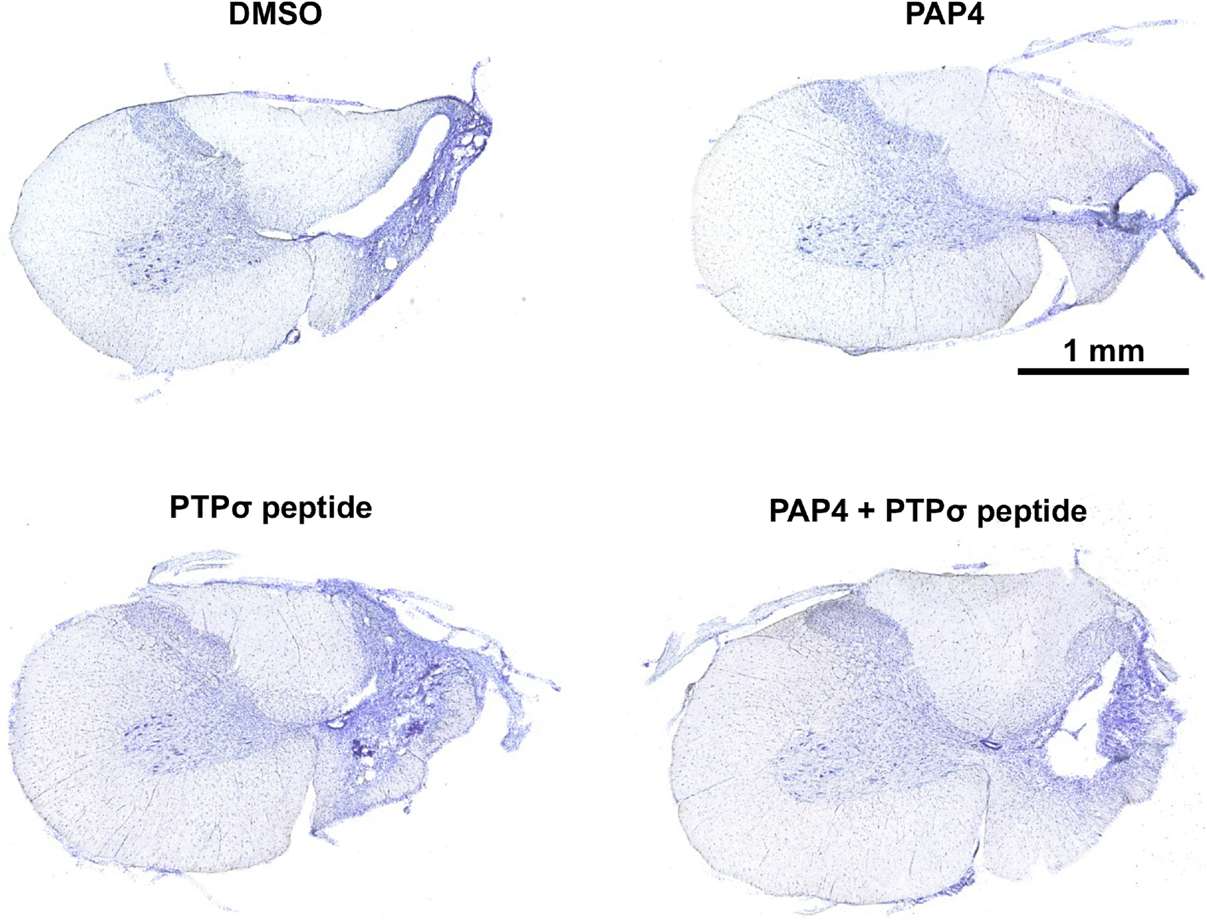
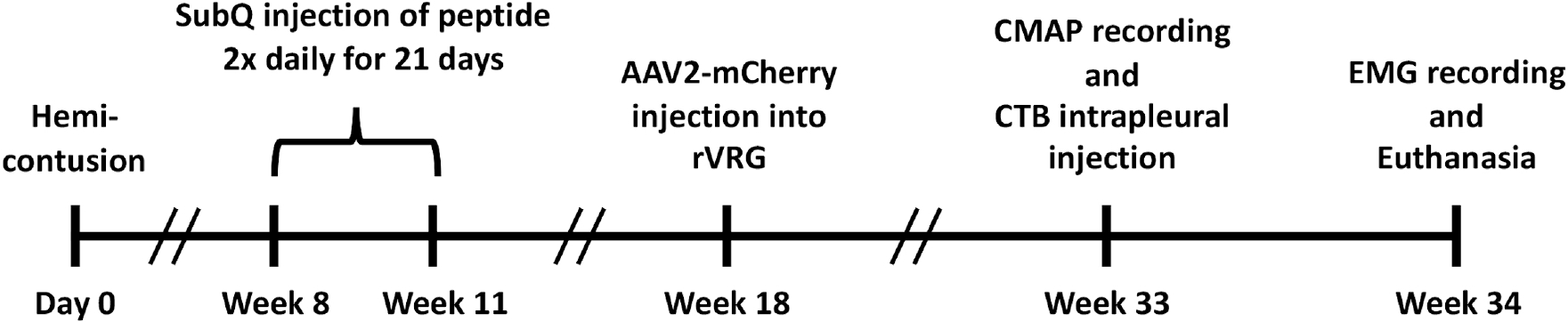
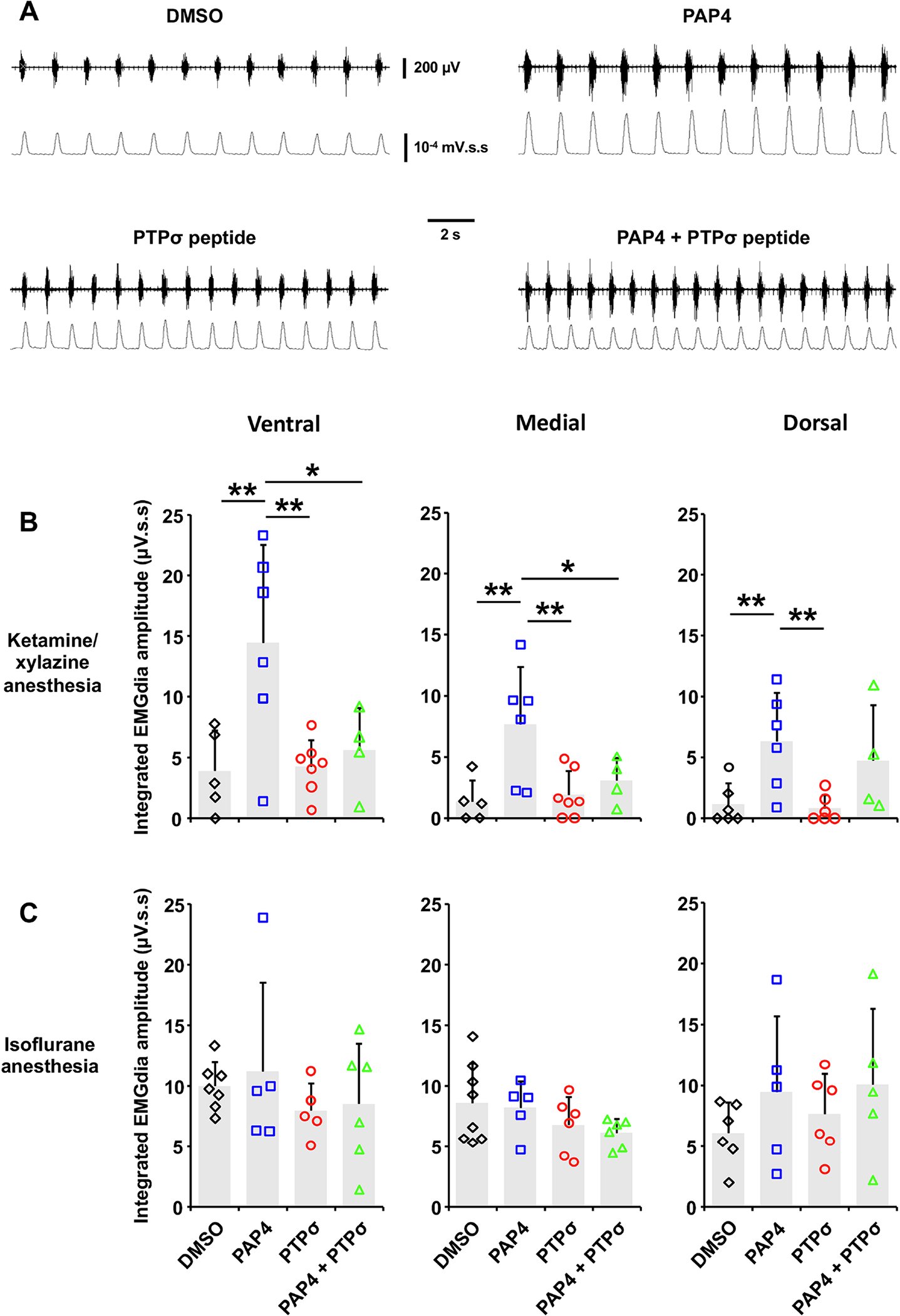
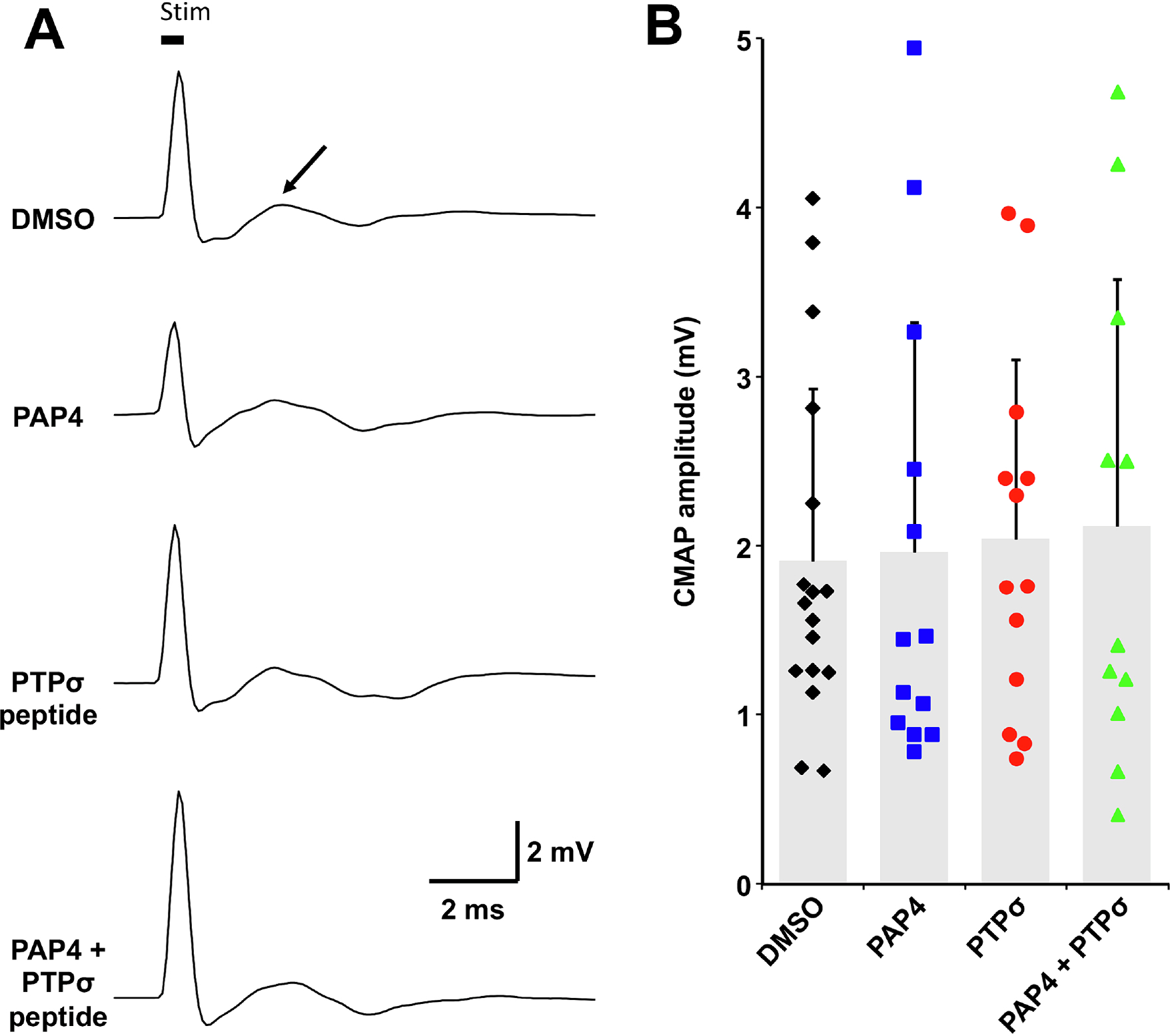
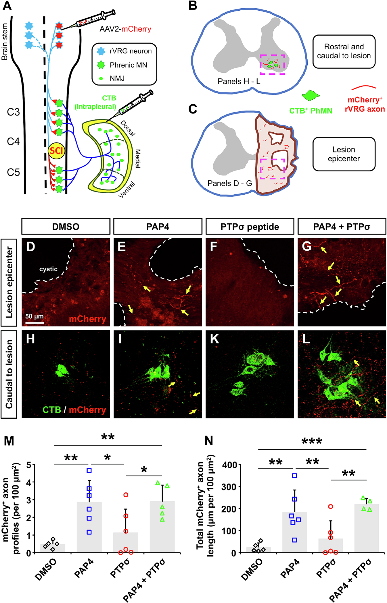
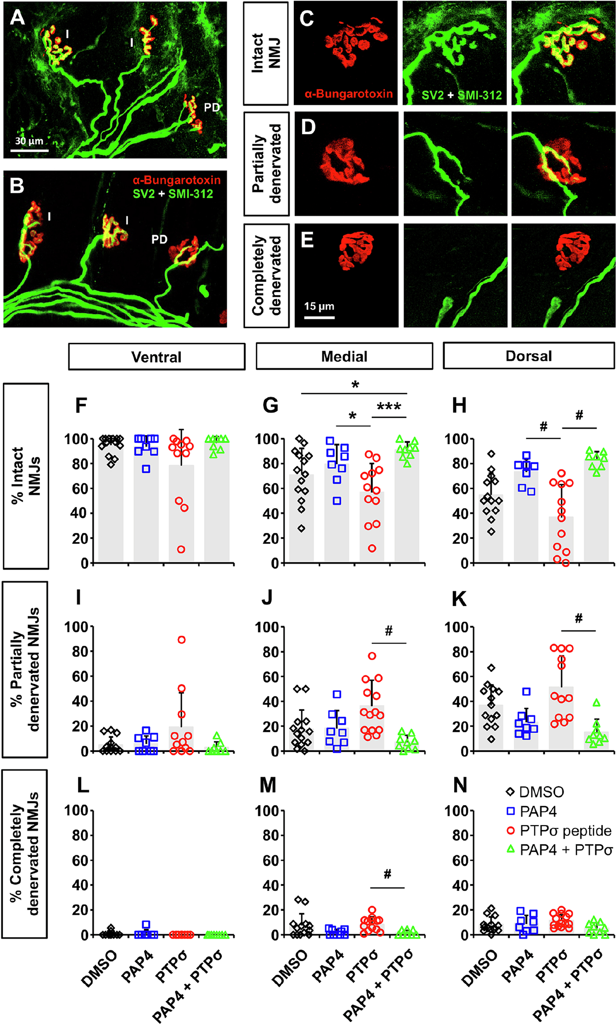
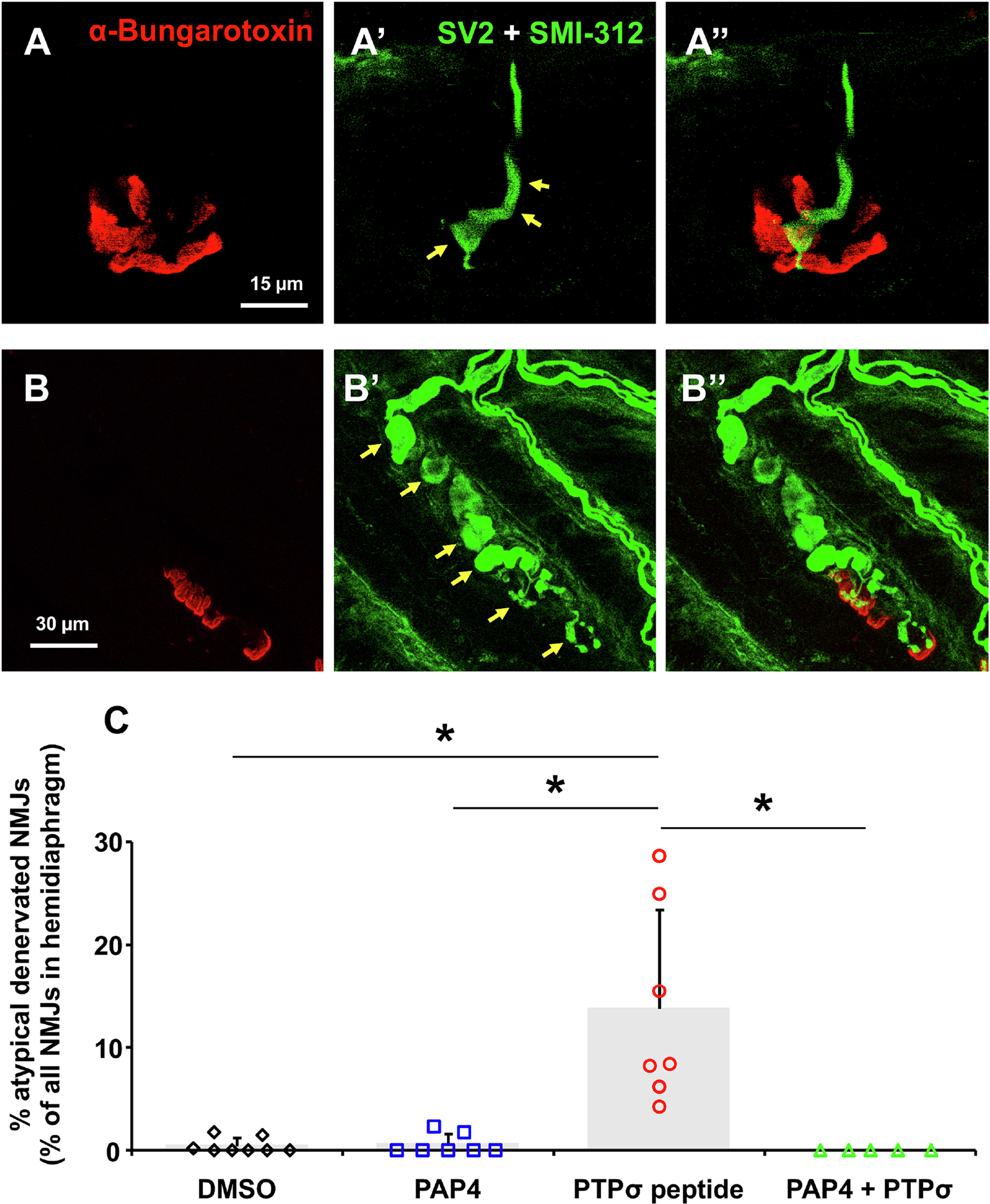
High spinal cord injury (SCI) leads to persistent and debilitating compromise in respiratory function. Cervical SCI not only causes the death of phrenic motor neurons (PhMNs) that innervate the diaphragm, but also damages descending respiratory pathways originating in the rostral ventral respiratory group (rVRG) located in the brainstem, resulting in denervation and consequent silencing of spared PhMNs located caudal to injury. It is imperative to determine whether interventions targeting rVRG axon growth and respiratory neural circuit reconnection are efficacious in chronic cervical contusion SCI, given that the vast majority of individuals are chronically-injured and most cases of SCI involve contusion-type damage to the cervical region. We therefore employed a rat model of chronic cervical hemicontusion to test therapeutic manipulations aimed at reconstructing damaged rVRG-PhMN-diaphragm circuitry to achieve recovery of respiratory function. At a chronic time point post-injury, we systemically administered: an antagonist peptide directed against phosphatase and tensin homolog (PTEN), a central inhibitor of neuron-intrinsic axon growth potential; an antagonist peptide directed against receptor-type protein tyrosine phosphatase sigma (PTPσ), another important negative regulator of axon growth capacity; or a combination of these two peptides. PTEN antagonist peptide (PAP4) promoted partial recovery of diaphragm motor activity out to nine months post-injury (though this effect depended on the anesthetic regimen used during recording), while PTPσ peptide did not impact diaphragm function after cervical SCI. Furthermore, PAP4 promoted robust growth of descending bulbospinal rVRG axons caudal to the injury within the denervated portion of the PhMN pool, while PTPσ peptide did not affect rVRG axon growth at this location that is critical to control of diaphragmatic respiratory function. In conclusion, we find that, when PTEN inhibition is targeted at a chronic time point following cervical contusion, our non-invasive PAP4 strategy can successfully promote significant regrowth of damaged respiratory neural circuitry and also partial recovery of diaphragm motor function.
Keywords: Axon; Breathing; Cervical; Chronic; Circuit; Contusion; Diaphragm; PTEN; PTPsigma; Regeneration; Respiratory; SCI; Spinal cord injury; Sprouting.
Copyright © 2024 Elsevier Inc. All rights reserved.
Conflict of interest statement
Declaration of competing interest The authors report no declarations of interest.
Figures
Update of
-
PTEN inhibition promotes robust growth of bulbospinal respiratory axons and partial recovery of diaphragm function in a chronic model of cervical contusion spinal cord injury.bioRxiv [Preprint]. 2024 Jan 11:2024.01.10.575021. doi: 10.1101/2024.01.10.575021. bioRxiv. 2024. Update in: Exp Neurol. 2024 Aug;378:114816. doi: 10.1016/j.expneurol.2024.114816. PMID: 38260313 Free PMC article. Updated. Preprint.
Similar articles
-
PTEN inhibition promotes robust growth of bulbospinal respiratory axons and partial recovery of diaphragm function in a chronic model of cervical contusion spinal cord injury.bioRxiv [Preprint]. 2024 Jan 11:2024.01.10.575021. doi: 10.1101/2024.01.10.575021. bioRxiv. 2024. Update in: Exp Neurol. 2024 Aug;378:114816. doi: 10.1016/j.expneurol.2024.114816. PMID: 38260313 Free PMC article. Updated. Preprint.
-
Protein Tyrosine Phosphatase σ Inhibitory Peptide Promotes Recovery of Diaphragm Function and Sprouting of Bulbospinal Respiratory Axons after Cervical Spinal Cord Injury.J Neurotrauma. 2020 Feb 1;37(3):572-579. doi: 10.1089/neu.2019.6586. Epub 2019 Sep 18. J Neurotrauma. 2020. PMID: 31392919 Free PMC article.
-
Respiratory axon regeneration in the chronically injured spinal cord.Neurobiol Dis. 2021 Jul;155:105389. doi: 10.1016/j.nbd.2021.105389. Epub 2021 May 8. Neurobiol Dis. 2021. PMID: 33975016 Free PMC article.
-
The Black Book of Psychotropic Dosing and Monitoring.Psychopharmacol Bull. 2024 Jul 8;54(3):8-59. Psychopharmacol Bull. 2024. PMID: 38993656 Free PMC article. Review.
-
An update on spinal cord injury and diaphragm neuromotor control.Expert Rev Respir Med. 2025 Jul;19(7):679-695. doi: 10.1080/17476348.2025.2495165. Epub 2025 Apr 22. Expert Rev Respir Med. 2025. PMID: 40258801 Review.
References
-
- Ahuja CS, Wilson JR, Nori S, Kotter MRN, Druschel C, et al. 2017. Traumatic spinal cord injury. Nat. Rev. Dis. Primers 3: 17018. - PubMed
-
- Arshadi C, Günther U, Eddison M, Harrington KIS, Ferreira TA. 2021. SNT: a unifying toolbox for quantification of neuronal anatomy. Nature Methods 18: 374–77 - PubMed
-
- Bhowmick S, Abdul-Muneer PM. 2020. PTEN Blocking Stimulates Corticospinal and Raphespinal Axonal Regeneration and Promotes Functional Recovery After Spinal Cord Injury. Journal of Neuropathology & Experimental Neurology 80: 169–81 - PubMed
MeSH terms
Substances
Grants and funding
LinkOut - more resources
Full Text Sources
Medical
Research Materials
