. 2024 Jun;39(3):501-512.
doi: 10.1016/j.virs.2024.05.006. Epub 2024 May 23.

Calmodulin-like 5 promotes PEDV replication by regulating late-endosome synthesis and innate immune response

Affiliations

Calmodulin-like 5 promotes PEDV replication by regulating late-endosome synthesis and innate immune response

Wen-Jun Tian et al. Virol Sin. 2024 Jun.

Abstract

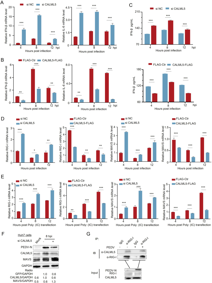
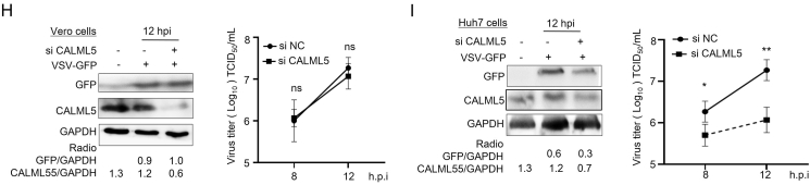
The infection caused by porcine epidemic diarrhea virus (PEDV) is associated with high mortality in piglets worldwide. Host factors involved in the efficient replication of PEDV, however, remain largely unknown. Our recent proteomic study in the virus-host interaction network revealed a significant increase in the accumulation of CALML5 (EF-hand protein calmodulin-like 5) following PEDV infection. A further study unveiled a biphasic increase of CALML5 in 2 and 12 ​h after viral infection. Similar trends were observed in the intestines of piglets in the early and late stages of the PEDV challenge. Moreover, CALML5 depletion reduced PEDV mRNA and protein levels, leading to a one-order-of-magnitude decrease in virus titer. At the early stage of PEDV infection, CALML5 affected the endosomal trafficking pathway by regulating the expression of endosomal sorting complex related cellular proteins. CALML5 depletion also suppressed IFN-β and IL-6 production in the PEDV-infected cells, thereby indicating its involvement in negatively regulating the innate immune response. Our study reveals the biological function of CALML5 in the virology field and offers new insights into the PEDV-host cell interaction.

Keywords: EF-hand protein calmodulin-like 5 (CALML5); Innate immune response; Late endosomes; Porcine epidemic diarrhea virus (PEDV).


Conflict of interest statement

Conflict of interest All authors declare that there are no competing interests.

Figures

Fig. 1
Fig. 1
The expression of CALML5 during PEDV infection both in vivo and in vitro. A, B The protein and mRNA levels of CALML5 during PEDV infection in Vero E6 cells. Cells were mock infected or infected with PEDV (CV777) at MOI of 1, and then harvested at pointed times. The protein level was analyzed by using Western blotting (WB) with the indicated antibodies (A). The mRNA level was subjected to RT-qPCR analysis for CALML5 (B). C, D Levels of CALML5 mRNA and proteins in jejunum of piglets during PEDV infection. Piglets were challenged with GIIb serotype PEDV isolated strain GS/2020/04 (105 TCID50 in 3 ​mL DMEM) or DMEM (3 ​mL) for different times, and the jejunum was obtained. The mRNA and protein levels of CALML5 were analyzed by RT-qPCR (C) and WB (D), respectively, in the tissue samples after cryogenic grinding with liquid nitrogen. Data presented as three independent experiments (means ​± ​SD). Statistical analyses were performed using one-way analysis of variance. ∗, P ​< ​0.05; ∗∗, P ​< ​0.01; ∗∗∗, P ​< ​0.001; ns showed no significant difference.
Fig. 2
Fig. 2
Effect of knockdown and overexpression of CALML5 on PEDV infection. AC Vero E6 cells were transfected with siRNA targeting CALML5 (siCALML5) or negative control (siNC), and then infected with PEDV (CV777) at an MOI of 1. Cells were harvested at pointed times. The protein level was analyzed by Western blotting (WB) (A), virus titer was determined using TCID50 with the method of Reed-Muench (B), and the mRNA level of PEDV-N was analyzed by RT-qPCR (C). D CALML5-knockout Vero cells and wild-type Vero cells were infected with 0.1 MOI PEDV (CV777), and then cells were harvested at pointed times. The protein level was analyzed by WB. E, F Vero cells were transfected with empty vector pFLAG or recombinant plasmid pFLAG-tagged CALML5. At 24 ​h post-transfection, cells were infected with PEDV (CV777) at an MOI of 0.1, and cells were harvested for indicated times. The protein level was analyzed by WB (E). Virus titer was determined using TCID50 with the method of Reed-Muench (F). Data presented as three independent experiments (means ​± ​SD). Statistical analyses were performed using one-way analysis of variance. ∗, P ​< ​0.05; ∗∗, P ​< ​0.01; ∗∗∗, P ​< ​0.001; ns showed no significant difference.
Fig. 3
Fig. 3
CALML5 is involved in viral internalization of PEDV. A, B CALML5 participates in PEDV internalization instead of viral attachment. Vero E6 cells were transfected with siCALML5 or siNC, and then infected with PEDV (CV777) at an MOI of 5 ​at 4 ​°C for 1 ​h. In viral attachment test (A), the infected cells were harvested after washing with PBS. In viral internalization test (B), after washing with PBS, cells were incubated at 37 ​°C for another 1 ​h. Then the infected cells were washed with PBS and harvested. The mRNA levels of PEDV-N analyzed by RT-qPCR. C Vero cells were pre-treated with rabbit antibody against CALML5 (diluted at 1:100) and DR5 (1:100) or rabbit IgG (1:100) for 2 ​h. After washing with PBS, cells were infected with PEDV (MOI of 5) at 37 ​°C for 2 ​h. The mRNA levels of PEDV-N analyzed by RT-qPCR. D CALML5 is also involved in PEDV release. Vero E6 cells transfected with siCALML5 or siNC and then infected with PEDV (CV777) at MOI of 0.5. PEDV titers in cells (intracellular) and culture fluid (extracellular) were determined by TCID50 assay at different infection times. E Vero E6 cells were transfected with siCALML5 or siNC and then infected with PEDV at MOI of 0.5 for different times, cell samples were collected, and the levels of negative-strand viral RNA (in the presence of CHX at 2 hpi) were analyzed by RT-qPCR. Data presented as three independent experiments (means ​± ​SD). Statistical analyses were performed using one-way analysis of variance. ∗, P ​< ​0.05; ∗∗, P ​< ​0.01; ∗∗∗, P ​< ​0.001; ns showed no significant difference.
Fig. 4
Fig. 4
CALML5 promotes the PEDV Infection by regulating late endosome in Vero cells. A, B Intracellular Ca2+ concentration quantified during PEDV infection. Vero E6 cells were transfected with siCALML5 or siNC (A), or Vero cells were pre-treated with DTZ (10 ​μmol/L) or DMSO (5 ​μL) for 2 ​h (B). After washing with PBS, cells were preloaded with Fluo-4AM and infected with PEDV at MOI of 1. The fluorescent absorbance values of cells were detected at different times with a fluorescent microplate reader. C Vero cells were pre-treated DTZ (10 ​μmol/L) or DMSO (5 ​μL), washed cells with PBS were challenged to PEDV (CV777) at the MOI of 1. The protein level was analyzed by Western blotting (WB) with the indicated antibodies. D Vero E6 cells were transfected with siCALML5 or siNC, and then mock infected or infected with PEDV at MOI of 1, the protein level was analyzed by WB with the indicated antibodies. E, F Late endosome inhibitor ABMA inhibits PEDV release. Vero cells pre-treated by ABMA (10 ​μmol/L) for 2 ​h, and then infected with PEDV at MOI of 0.1 for 24 ​h. The protein level was analyzed by WB with the indicated antibodies (E) and PEDV titers in cells (intracellular) and culture fluid (extracellular) were determined by TCID50 assay at different infection times (F). G Immunofluorescence assay. The Vero cells were infected with PEDV at MOI of 1, and were then fixed at pointed times and incubated with mouse monoclonal antibody anti-TSG101 (top) and mouse monoclonal antibody anti-EEA1 (bottom), followed by FITC-conjugated goat anti-mouse secondary antibody (green). Cells were then incubated with rabbit anti-CALML5 followed by TRITC-conjugated goat anti-mouse secondary antibody. Finally, the cells were counterstained with DAPI (blue), and the images were viewed under an ECLIPSE Ni series upright biological microscope (Fluoview 200×). Data presented as three independent experiments (means ​± ​SD). Statistical analyses were performed using one-way analysis of variance. ∗, P ​< ​0.05; ∗∗, P ​< ​0.01.
Fig. 5
Fig. 5
CALML5 regulates late endosome traffic by targeting ESCRT. A Vero E6 cells were transfected with siCALML5 or siNC, and mRNA levels of ESCRT components were analyzed by RT-qPCR. B Quantitative analysis in mRNA level of ESCRT components. Vero E6 cells were transfected with siCALML5 or siNC, and then mock infected or infected with PEDV at MOI of 1 for indicated times. The mRNA level of ESCRT components was analyzed by RT-qPCR. C The effect of knockdown of ESCRT components on PEDV replication. Vero cells were transfected with siTSG101, siVPS28, siMVB12, siEAP20 or siNC, and then infected with PEDV (CV777) at MOI of 1 for 12 ​h (left) or 24 ​h (right). The PEDV-NP level was quantified by Western blotting (WB) and the intensity band ratio of PEDV-NP to GAPDH was analyzed by using ImageJ software (B). Under the same experimental conditions, titers of viruses were measured, as log10 ​PFU/mL (C). DF A screened ESCRT components participate in viral internalization of PEDV. Vero E6 cells were transfected with siTSG101, siVPS28, siMVB12, siEAP20 or siNC, and infected with PEDV (CV777) at an MOI of 5 ​at 4 ​°C for 1 ​h. In viral attachment test (D, left), the infected cells were harvested after washing with PBS. In viral internalization test (D, right), after washing with PBS, cells were incubated at 37 ​°C for another 1 ​h. Then the infected cells were washed with PBS and harvested. The mRNA levels of PEDV-N were analyzed by RT-qPCR. E A screened ESCRT components are involved in PEDV release. Vero cells transfected with siTSG101, siVPS28, siMVB12, siEAP20 or siNC, and then infected with PEDV at MOI of 0.5 for 12 ​h. PEDV titers in cells (intracellular) and culture fluid (extracellular) were determined by TCID50 assay at different times. F, G Cytotoxicity of the siRNAs and knockdown efficiency. The Vero cells grown in 96-well plates were transfected with the indicated siRNAs, and the cell viability was evaluated by using CCK8 method (F). Total RNA was extracted and mRNA level was analyzed by RT-qPCR (G). The data were presented as mean ​± ​SEM of three independent experiments. Statistical analyses were performed using one-way analysis of variance. ∗, P ​< ​0.05; ∗∗, P ​< ​0.01; ∗∗∗, P ​< ​0.001; ns showed no significant difference.
Fig. 6
Fig. 6
CALML5 is involved in PEDV replication in Huh-7 ​cells. A The protein expression of CALML5 during PEDV infection in Huh-7 ​cells. Huh-7 ​cells were mock infected or infected with PEDV (CV777) at MOI of 1, cells were harvested at pointed times. The protein level was analyzed by Western blotting (WB). B, C Huh-7 ​cells were transfected with siCALML5 or siNC, and then infected with PEDV at an MOI of 1, and cells were harvested at pointed times. The protein level was analyzed by WB (B), and virus titer was determined using TCID50 with the method of Reed-Muench (C). D Late-endosome inhibitor ABMA was treated in Huh-7 ​cells. Huh-7 ​cells were pre-treated with 10 ​μmol/L ABMA for 2 ​h, and then infected with PEDV at MOI of 1 for 24 ​h. The protein level was analyzed by WB with the indicated antibodies. The data were presented as mean ​± ​SEM of three independent experiments. Statistical analyses were performed using one-way analysis of variance. ∗, P ​< ​0.05; ∗∗, P ​< ​0.01.
Fig. 7
Fig. 7
CALML5 negatively regulates the innate immunity by targeting RIG-I-MAVS pathway. AD In knockdown group, Huh-7 ​cells were transfected with siCALML5 or siNC, and then infected with PEDV (CV777) at an MOI of 1, and cells were harvested at pointed times. The mRNA level of IFN-β (A, left), IL-6 (A, right), RIG-I (D, left), and MAVS (D, right) was analyzed by RT-qPCR. In overexpression group, Huh-7 ​cells were transfected with empty vector pFLAG or recombinant plasmid pFLAG-CALML5 for 24 ​h, cells were infected with PEDV at an MOI of 0.1, and cells were harvested at pointed times. The mRNA level of IFN-β (B, left), IL-6 (B, right), RIG-I (D, left), and MAVS (D, right) was analyzed by RT-qPCR. The production of endogenous IFN-β was quantified by ELISA kit (C). E Huh-7 ​cells were treated as above and then transfected with poly (I:C) for 6 ​h. The cells were harvested at pointed times. The mRNA level of RIG-I and MAVS was analyzed by RT-qPCR. F Huh-7 ​cells were transfected with siCALML5 or siNC, then infected with PEDV at an MOI of 1 for 8 ​h. The protein level was analyzed by Western blotting (WB) with the indicated antibodies. G Co-immunoprecipitation (co-IP) assay of CALML5 and RIG-I. Huh-7 ​cells were mock infected or infected with PEDV at MOI of 0.1 for 12 ​h, and cell lysates were generated for co-IP assays using immunoglobulin G (IgG) or RIG-I antibody (α-RIG-I), followed by WB to analyze levels of RIG-I and CALML5. H, I Vero cells (H) or Huh 7 ​cells (I) were transfected with siCALML5 or siNC, then infected with GFP-VSV at a MOI of 0.1 for 12 ​h, cell lysates were analyzed by WB with specific antibodies to detect CALML5 and VSV-G, with GAPDH as a control (left). Under the same experimental condition, titers of viruses were measured, as log10 ​PFU/mL (right). Values represent means of triplicates with SD. ∗P ​< ​0.05, ∗∗P ​< ​0.01; ∗∗∗P ​< ​0.001; ns showed no significant difference.
Fig. 7
Fig. 7
CALML5 negatively regulates the innate immunity by targeting RIG-I-MAVS pathway. AD In knockdown group, Huh-7 ​cells were transfected with siCALML5 or siNC, and then infected with PEDV (CV777) at an MOI of 1, and cells were harvested at pointed times. The mRNA level of IFN-β (A, left), IL-6 (A, right), RIG-I (D, left), and MAVS (D, right) was analyzed by RT-qPCR. In overexpression group, Huh-7 ​cells were transfected with empty vector pFLAG or recombinant plasmid pFLAG-CALML5 for 24 ​h, cells were infected with PEDV at an MOI of 0.1, and cells were harvested at pointed times. The mRNA level of IFN-β (B, left), IL-6 (B, right), RIG-I (D, left), and MAVS (D, right) was analyzed by RT-qPCR. The production of endogenous IFN-β was quantified by ELISA kit (C). E Huh-7 ​cells were treated as above and then transfected with poly (I:C) for 6 ​h. The cells were harvested at pointed times. The mRNA level of RIG-I and MAVS was analyzed by RT-qPCR. F Huh-7 ​cells were transfected with siCALML5 or siNC, then infected with PEDV at an MOI of 1 for 8 ​h. The protein level was analyzed by Western blotting (WB) with the indicated antibodies. G Co-immunoprecipitation (co-IP) assay of CALML5 and RIG-I. Huh-7 ​cells were mock infected or infected with PEDV at MOI of 0.1 for 12 ​h, and cell lysates were generated for co-IP assays using immunoglobulin G (IgG) or RIG-I antibody (α-RIG-I), followed by WB to analyze levels of RIG-I and CALML5. H, I Vero cells (H) or Huh 7 ​cells (I) were transfected with siCALML5 or siNC, then infected with GFP-VSV at a MOI of 0.1 for 12 ​h, cell lysates were analyzed by WB with specific antibodies to detect CALML5 and VSV-G, with GAPDH as a control (left). Under the same experimental condition, titers of viruses were measured, as log10 ​PFU/mL (right). Values represent means of triplicates with SD. ∗P ​< ​0.05, ∗∗P ​< ​0.01; ∗∗∗P ​< ​0.001; ns showed no significant difference.
Fig. 8
Fig. 8
Schematic diagram of CALML5 promotes PEDV replication by regulating late-endosome synthesis and innate immune response. CALML5 promotes PEDV virion internalization to influence late-endosome synthesis by regulating ESCRT in Vero E6 cells (left). Right, CALML5 suppresses the production of IFN-β by targeting RIG-I-MAVS pathway in Huh-7 ​cells.

Similar articles

References

    1. Bai D., Fang L., Xia S., Ke W., Wang J., Wu X., Fang P., Xiao S. Porcine deltacoronavirus (PDCoV) modulates calcium influx to favor viral replication. Virology. 2020;539:38–48. - PMC - PubMed
    1. Barouch-Bentov R., Neveu G., Xiao F., Beer M., Bekerman E., Schor S., Campbell J., Boonyaratanakornkit J., Lindenbach B., Lu A., Jacob Y., Einav S. Hepatitis C virus proteins interact with the endosomal sorting complex required for transport (ESCRT) machinery via ubiquitination to facilitate viral envelopment. mBio. 2016;7 - PMC - PubMed
    1. Bu J., Zhang Y., Niu N., Bi K., Sun L., Qiao X., Wang Y., Zhang Y., Jiang X., Wang D., Ma Q., Li H., Liu C. Dalpiciclib partially abrogates ER signaling activation induced by pyrotinib in HER2(+)HR(+) breast cancer. Elife. 2023;12 - PMC - PubMed
    1. Deng L., Liang Y., Ariffianto A., Matsui C., Abe T., Muramatsu M., Wakita T., Maki M., Shibata H., Shoji I. Hepatitis C virus-induced ROS/JNK signaling pathway activates the E3 ubiquitin ligase itch to promote the release of HCV particles via polyubiquitylation of VPS4A. J. Virol. 2022;96 - PMC - PubMed
    1. Dong H.J., Wang J., Zhang X.Z., Li C.C., Liu J.F., Wang X.J. Proteomic screening identifies RPLp2 as a specific regulator for the translation of coronavirus. Int. J. Biol. Macromol. 2023;230 - PMC - PubMed

MeSH terms

LinkOut - more resources