. 2024 Jun;39(3):469-477.
doi: 10.1016/j.virs.2024.05.007. Epub 2024 May 22.

Function investigation of p11.5 in ASFV infection

Affiliations

Function investigation of p11.5 in ASFV infection

Dan Yin et al. Virol Sin. 2024 Jun.

Abstract

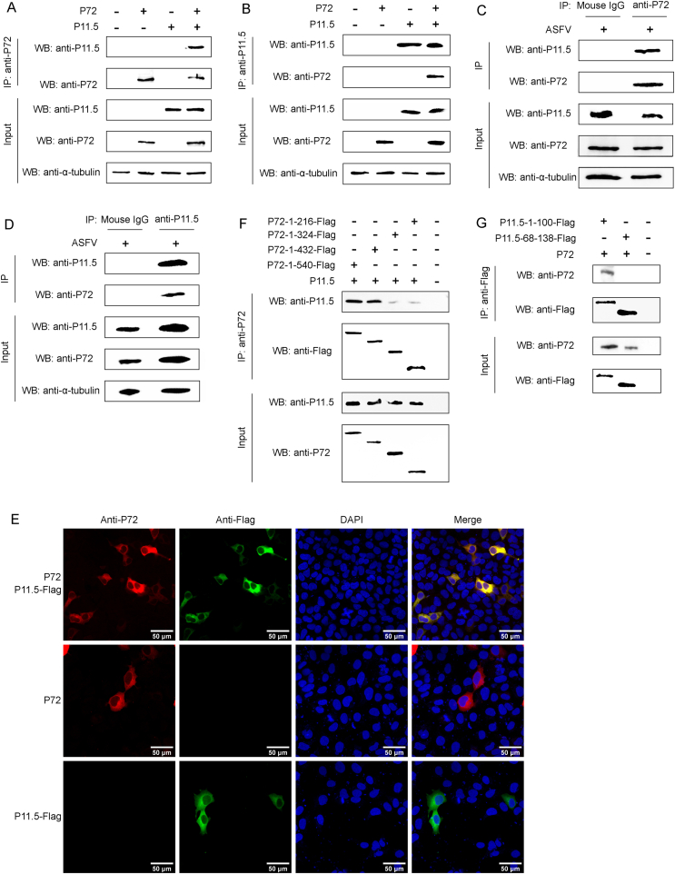
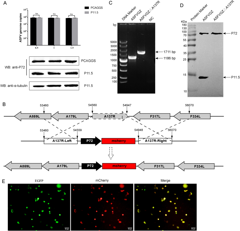
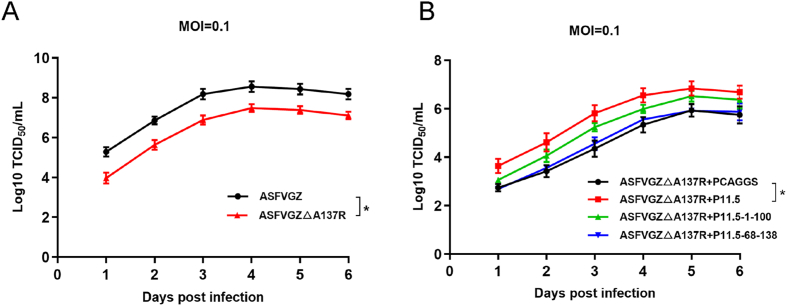
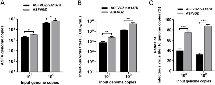
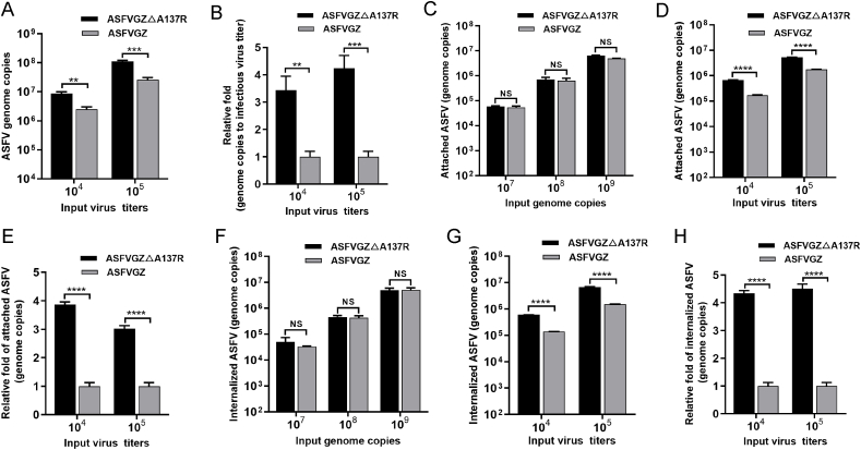
Virus replication relies on complex interactions between viral proteins. In the case of African swine fever virus (ASFV), only a few such interactions have been identified so far. In this study, we demonstrate that ASFV protein p72 interacts with p11.5 using co-immunoprecipitation and liquid chromatography-mass spectrometry (LC-MS). It was found that protein p72 interacts specifically with p11.5 ​at sites amino acids (aa) 1-216 of p72 and aa 1-68 of p11.5. To assess the importance of p11.5 in ASFV infection, we developed a recombinant virus (ASFVGZΔA137R) by deleting the A137R gene from the ASFVGZ genome. Compared with ASFVGZ, the infectious progeny virus titers of ASFVGZΔA137R were reduced by approximately 1.0 logs. In addition, we demonstrated that the growth defect was partially attributable to a higher genome copies-to-infectious virus titer ratios produced in ASFVGZΔA137R-infected MA104 ​cells than in those infected with ASFVGZ. This finding suggests that MA104 ​cells infected with ASFVGZΔA137R may generate larger quantities of noninfectious particles. Importantly, we found that p11.5 did not affect virus-cell binding or endocytosis. Collectively, we show for the first time the interaction between ASFV p72 and p11.5. Our results effectively provide the relevant information of the p11.5 protein. These results extend our understanding of complex interactions between viral proteins, paving the way for further studies of the potential mechanisms and pathogenesis of ASFV infection.

Keywords: African swine fever virus; Protein interactions; p11.5 protein; p72 protein.


Conflict of interest statement

Conflict of interest All authors declare that there are no competing interests.

Figures

Fig. 1
Fig. 1
Silver staining was used to detect the enrichment of p72 interacting proteins. A Dynamic growth of ASFVGZ in IPAM and PK-15 ​cells in vitro. IPAM or PK-15 ​cells were infected (MOI ​= ​0.1) with ASFVGZ and viral genome copies were calculated at the indicated times post-infection. PK-15 ​cells (B) and IPAM cells (C) were infected with ASFVGZ at 0.1 MOI. Anti-p72 mAb were used for CO-IP 48 ​h after cell infection. p72-interacting proteins were eluted with protein G sepharose and analyzed on SDS-PAGE followed by silver staining. The normal mouse IgG as a negative control. The data were tested three times independently. Arrows indicate differential bands.
Fig. 2
Fig. 2
Protein p11.5 interacts with p72. A-B HEK293T cells were co-transfected with the pCAGGS-p72 and pCAGGS-p11.5 expression plasmids for 48 ​h, followed by a CO-IP assay for p11.5 protein and p72 protein using anti-p72 (A) or anti-p11.5 (B) antibody. C-D MA104 ​cells were infected with ASFVGZ at an MOI of 0.1, and cell extracts were analyzed by Co-IP at 48 ​h post infection using anti-p72 antibody (C) or anti-p11.5 (D) antibody. (E) MA104 ​cells were transfected with pCAGGS-p72 and/or pCAGGS-p11.5-Flag expression plasmids for 24 ​h and then fixed and processed for dual labeling. p11.5 (green) and p72 (red) proteins were visualized by immunostaining with rabbit anti-Flag and mouse anti-p72 antibodies. Cell nuclei were counterstained with DAPI (blue). The areas of colocalization in merged images are shown in yellow. (F) The truncated p72 and full-length p11.5 were co-transfected into HEK293T cells. After 48 ​h, the cell lysates were immunoprecipitated with anti-p72 and analyzed by WB. (G) The truncated p11.5 and full-length p72 were co-transfected into HEK293T cells. After 48 ​h, the cell lysates were immunoprecipitated with anti-Flag and analyzed by WB.
Fig. 3
Fig. 3
Construction of an ASFVGZΔA137R deletion mutant. A MA104 ​cells were transfected with pCAGGS-p11.5 for 24 ​h and inoculated with ASFVGZ at an MOI of 0.1. The gene and protein expression levels of p72 were quantified by qPCR and Western blot. B Schematic diagram for the construction of ASFVGZΔA137R. The open reading frame of A137R was replaced by an mCherry expression cassette as shown. C PCR analysis of ASFVGZ and ASFVGZΔA137R. NC, negative control. D Detection of p11.5 expression by Western blot. E MA104 ​cells were infected with the recombinant ASFVGZΔA137R deletion mutant showing EGFP and mCherry expression.
Fig. 4
Fig. 4
Kinetics of growth of ASFVGZΔA137R and parental ASFVGZ in MA104 ​cells in vitro. A MA104 ​cells were infected (MOI ​= ​0.1) with each of the viruses, and virus yields were titrated with MA104 ​cells at the indicated times post-infection. B MA104 ​cells were transfected with pCAGGS-p11.5, pCAGGS-p11.5-1-100, pCAGGS-p11.5-68-138 or pCAGGS empty vector respectively and then infected with ASFVGZΔA137R at 0.1 MOI. The virus yields were titrated with MA104 ​cells at the indicated times post-infection. Data represent means and SD from three independent experiments. The significance of differences between groups was determined using Student's t-test (∗P ​< ​0.05).
Fig. 5
Fig. 5
ASFVGZΔA137R produces a reduced ratio of infectious virus titer-to-genome copies in MA104 ​cells. A Genome copies of progeny ASFVGZΔA137R or ASFVGZ. MA104 cells in 24-well plates were infected with either ASFVGZΔA137R or ASFVGZ at genome copies of 108 or 109. The genome copies in supernatant were determined by qPCR at 24 hpi. B Infectious progeny virus production of ASFVGZΔA137R or ASFVGZ. The titers of infectious progeny virions from group A were detected according to the Spearman–Kärber method. C ASFVGZΔA137R or ASFVGZ infectious virus titer-to-genome copies ratios. The infectious virus titer-to-genome copies ratios were calculated based on the ratio of (B) to the (A) at 24 hpi (n ​= ​3). The significance of differences between groups was determined using Student's t-test (∗P ​< ​0.05; ∗∗P ​< ​0.01; ∗∗∗P ​< ​0.001).
Fig. 6
Fig. 6
The p11.5 protein is not required for ASFV binding to or entry into MA104 ​cells. A Genome copies of ASFVGZΔA137R or ASFVGZ per 104 and 105 TCID50. B ASFVGZΔA137R or ASFVGZ genome copies-to-infectious virus titer ratios based on samples from panel A. C-D The attachment levels of ASFVGZΔA137R or ASFVGZ were similar. Equal numbers of genome copies (107, 108, and 109) (C) or equal titers (104 and 105) (D) of ASFVGZΔA137R or ASFVGZ were added to MA104 ​cells at 4 ​°C and allowed to attach for 2 ​h. The numbers of genome copies of attached ASFVs were quantified by qPCR. E The fold change in attached ASFVGZΔA137R was nearly 4 times higher than that of ASFVGZ with equal titers (D). F-G The internalization levels of ASFVGZΔA137R or ASFVGZ were similar. Equal numbers of genome copies (107, 108, and 109) (F) or equal titers (104 and 105) (G) of ASFVGZΔA137R or ASFVGZ were added to MA104 ​cells at 37 ​°C and allowed to internalize for 2 ​h. The genome copies of internalized ASFVs were quantified by qPCR. H The fold change in internalized ASFVGZΔA137R was approximately 4 times higher than that of ASFVGZ with equal titers (G). The data shown are from three independent experiments. The significance of differences between groups was determined using Student's t-test (∗∗P ​< ​0.01; ∗∗∗P ​< ​0.001; ∗∗∗∗P ​< ​0.0001; NS, not significant).

Similar articles

Cited by

References

    1. Alcami A., Angulo A., Vinuela E. Mapping and sequence of the gene encoding the African swine fever virion protein of M(r) 11500. J. Gen. Virol. 1993;74:2317–2324. - PubMed
    1. Alejo A., Matamoros T., Guerra M., Andres G. A proteomic atlas of the African swine fever virus particle. J. Virol. 2018;92 18. - PMC - PubMed
    1. Andres G., Charro D., Matamoros T., Dillard R.S., Abrescia N.G.A. The cryo-EM structure of African swine fever virus unravels a unique architecture comprising two icosahedral protein capsids and two lipoprotein membranes. J. Biol. Chem. 2020;295:1–12. - PMC - PubMed
    1. Andres G., Garcia-Escudero R., Vinuela E., Salas M.L., Rodriguez J.M. African swine fever virus structural protein pE120R is essential for virus transport from assembly sites to plasma membrane but not for infectivity. J. Virol. 2001;75:6758–6768. - PMC - PubMed
    1. Borca M.V., Ramirez-Medina E., Silva E., Vuono E., Rai A., Pruitt S., Holinka L.G., Velazquez-Salinas L., Zhu J., Gladue D.P. Development of a highly effective African swine fever virus vaccine by deletion of the I177L gene results in sterile immunity against the current epidemic Eurasia strain. J. Virol. 2020;94 19. - PMC - PubMed

LinkOut - more resources