. 2024 Sep-Oct;99(5):696-705.
doi: 10.1016/j.abd.2023.11.005. Epub 2024 May 23.

Role of tangential biopsy in the diagnosis of nail psoriasis

Affiliations

Role of tangential biopsy in the diagnosis of nail psoriasis

Laura Bertanha et al. An Bras Dermatol. 2024 Sep-Oct.

Abstract

Background: Histopathology can be crucial for diagnosis of inflammatory nail diseases. Longitudinal excision and punch biopsies are the most used techniques to obtain the tissue sample. However, there is a low clinical-histopathological correlation, besides the risk of nail dystrophy. Tangential excision biopsy (TB) is a well-established technique for the investigation of longitudinal melanonychia. TB could also be used to evaluate diseases in which histopathological changes are superficial, as in psoriasis.

Objective: To study the value of TB in the histopathological diagnosis of nail psoriasis.

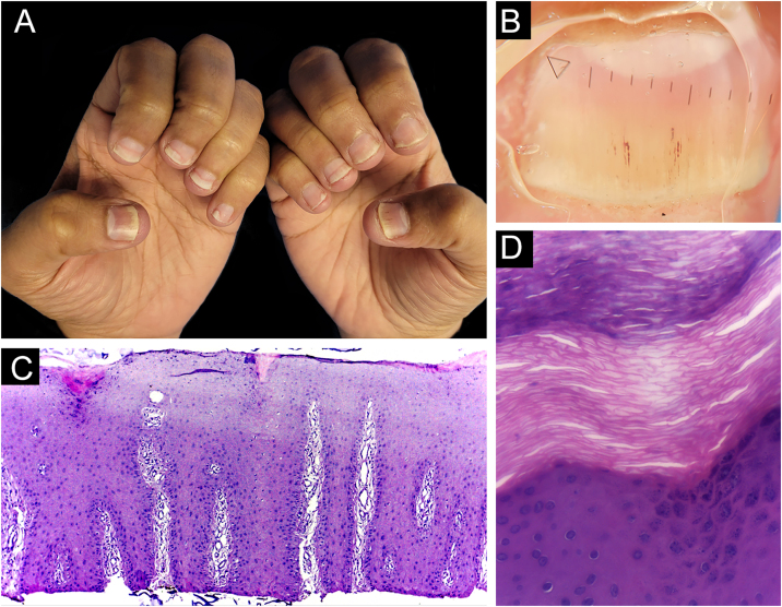
Methods: This is a prospective and descriptive study of the clinical-histopathological findings of samples from the nail bed or matrix and nail plate of 13 patients with clinical suspicion of nail psoriasis. Biopsies were obtained through partial nail avulsion and TB.

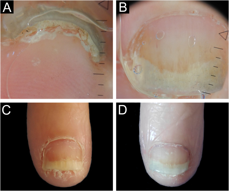
Results: In nine patients, the hypothesis of psoriasis was confirmed by histopathology; in one, the criteria for diagnosing nail lichen planus were fulfilled. The tissue sample of only one patient did not reach the dermal papillae, and, in four of 13 patients, the adventitial dermis was not sampled. No patient developed onychodystrophy after the procedure.

Study limitations: In three patients, the clinical and, consequently, histopathological nail changes were subtle. Also, in one patient's TB didn't sample the dermal papillae.

Conclusions: TB is a good option to assist in the histopathological diagnosis of nail psoriasis, especially when appropriate clinical elements are combined. Using this technique, larger and thinner samples, short postoperative recovery time, and low risk of onychodystrophy are obtained.

Keywords: Biopsy; Histopathology; Nail diseases; Psoriasis.


Figures

Figure 1
Figure 1
Longitudinal tangential biopsy technique – (A) Onychoscopy: regular onycholysis with salmon patch and dilated linear vessels; (B) Partial detachment of the nail plate with a number 15 scalpel blade, in the demarcated region; (C) Partial cutting of the plate with pliers; (D) Slightly thicker tangential biopsy of the nail bed (sample measuring 0.7 × 0.2 × 0.1 cm).
Figure 2
Figure 2
Lichen planus ‒ (A) All fingernails affected: regular onycholysis, subungual hyperkeratosis and onychodystrophy; (B) Onychoscopy: red spots on the lunula, subungual hyperkeratosis and irregular striated leukonychia; (C) Nail bed with significant lymphoid infiltrate within the dermal papillae; (D) lichenoid lymphoid infiltrate fills the papilla, with a subepidermal cleft. Hematoxilina & eosin, ×100 (C) and × 400 (D).
Figure 3
Figure 3
Nail psoriasis ‒ (A) Regular onycholysis with salmon patch on several digits; (B) Onychoscopy: regular onycholysis with salmon patch; (C) Coarse nail pittings.
Figure 4
Figure 4
Nail psoriasis ‒ (A) Regular onycholysis with salmon patch on several digits; (B) Compact subungual hyperkeratosis, seen on the free nail edge; (C) Onychoscopy: regular onycholysis with salmon patch and punctate leukonychia.
Figure 5
Figure 5
Psoriasis (nail bed) ‒ (A) Psoriasiform hyperplasia of the epidermis, hypervascularized papillary dermis and thinning of the suprapapillary stratum spinosum; (B and C) Thinning of the suprapapillary stratum spinosum, highly vascularized papillae and absence of spongiosis; (D) Parakeratosis and intracorneal neutrophils exudate. Hematoxilina & eosin, 40 (A) and ×400 (B‒D).
Figure 6
Figure 6
Psoriasis (nail plate) ‒ (A) Papillomatosis (the tops of the papillae superficialized from the bed – red arrows); (B) Intracorneal serous exudate on the top of the papillae (blue arrows), with parakeratosis (black arrows); (C) Keratinocyte nuclei (parakeratosis ‒ black arrows) and rare neutrophils (yellow arrows). Hematoxilina & eosin, ×100 (A, B) and ×400 (C).
Figure 7
Figure 7
Nail psoriasis -before and after tangential biopsy ‒ (A) Onychoscopy of the area to be biopsied- regular onycholysis with salmon patches and splinter hemorrhages; (B) 7-day postoperative period ‒ excellent bed healing; (C) 30-days post-operative period – complete nail recovery.
Figure 8
Figure 8
Nail psoriasis -before and after tangential biopsy ‒ (A and B) Onychoscopy of the area to be biopsied - regular onycholysis with oil spot, splinter hemorrhages and compact subungual hyperkeratosis; (C) Immediate pre-operative period; (D) 30-days post-operative – complete nail recovery.
Figure 9
Figure 9
Nail psoriasis ‒ (A) All fingernails with regular onycholysis, salmon patch, some oil spots, subungual hyperkeratosis and splinter hemorrhages; (B) Onychoscopy: distal leukonychia and splinter hemorrhages; (C) High papillae vascular density; (D) Area of hypogranulosis, on the left, and area of hypergranulosis, on the right. Hematoxilina & eosin ×100 (C) and ×400 (D).
Figure 10
Figure 10
Nail psoriasis ‒ (A) Regular onycholysis on all fingernails, some with oil spots, subungual hyperkeratosis, and total dystrophy; (B) Neutrophilis exudate amidst superficial keratinocytes (red arrow); neutrophils within parakeratotic scale (yellow arrows). Hematoxilina & eosin, ×400.

References

    1. Grover C., Chaturvedi U.K., Reddy B.S. Role of nail biopsy as a diagnostic tool. Indian J Dermatol Venereol Leprol. 2012;78:290–298. - PubMed
    1. Schons K.R.R., Knob C.F., Murussi N., Beber A.A.C., Neumaier W., Monticielo O.A. Nail psoriasis: a review of the literature. An Bras Dermatol. 2014;89:312–317. - PMC - PubMed
    1. Farias D.C., Tosti A., Chiacchio N.D., Hirata S.H. Dermoscopy in nail psoriasis. An Bras Dermatol. 2010;85:101–103. - PubMed
    1. Tan E.S., Chong W.S., Tey H.L. Nail psoriasis: a review. Am J Clin Dermatol. 2012;13:375–388. - PubMed
    1. Hanno R., Mathes B.M., Krull E.A. Longitudinal nail biopsy in evaluation of acquired nail dystrophies. J Am Acad Dermatol. 1986;14:803–809. - PubMed

LinkOut - more resources