. 2024 Jul;48(7):1289-1301.
doi: 10.1111/acer.15350. Epub 2024 May 24.

Prenatal ethanol and cannabis exposure have sex- and region-specific effects on somatostatin and neuropeptide Y interneurons in the rat hippocampus

Affiliations

Prenatal ethanol and cannabis exposure have sex- and region-specific effects on somatostatin and neuropeptide Y interneurons in the rat hippocampus

Hannah M O Reid et al. Alcohol Clin Exp Res (Hoboken). 2024 Jul.

Abstract

Background: Cannabis is increasingly being legalized and socially accepted around the world and is often used with alcohol in social settings. We recently showed that in utero exposure to both substances can alter the density of parvalbumin-expressing interneurons in the hippocampus. Here we investigate the effects of in utero alcohol and cannabis exposure, alone or in combination, on somatostatin- and neuropeptide Y-positive (NPY) interneurons. These are separate classes of interneurons important for network synchrony and inhibition in the hippocampus.

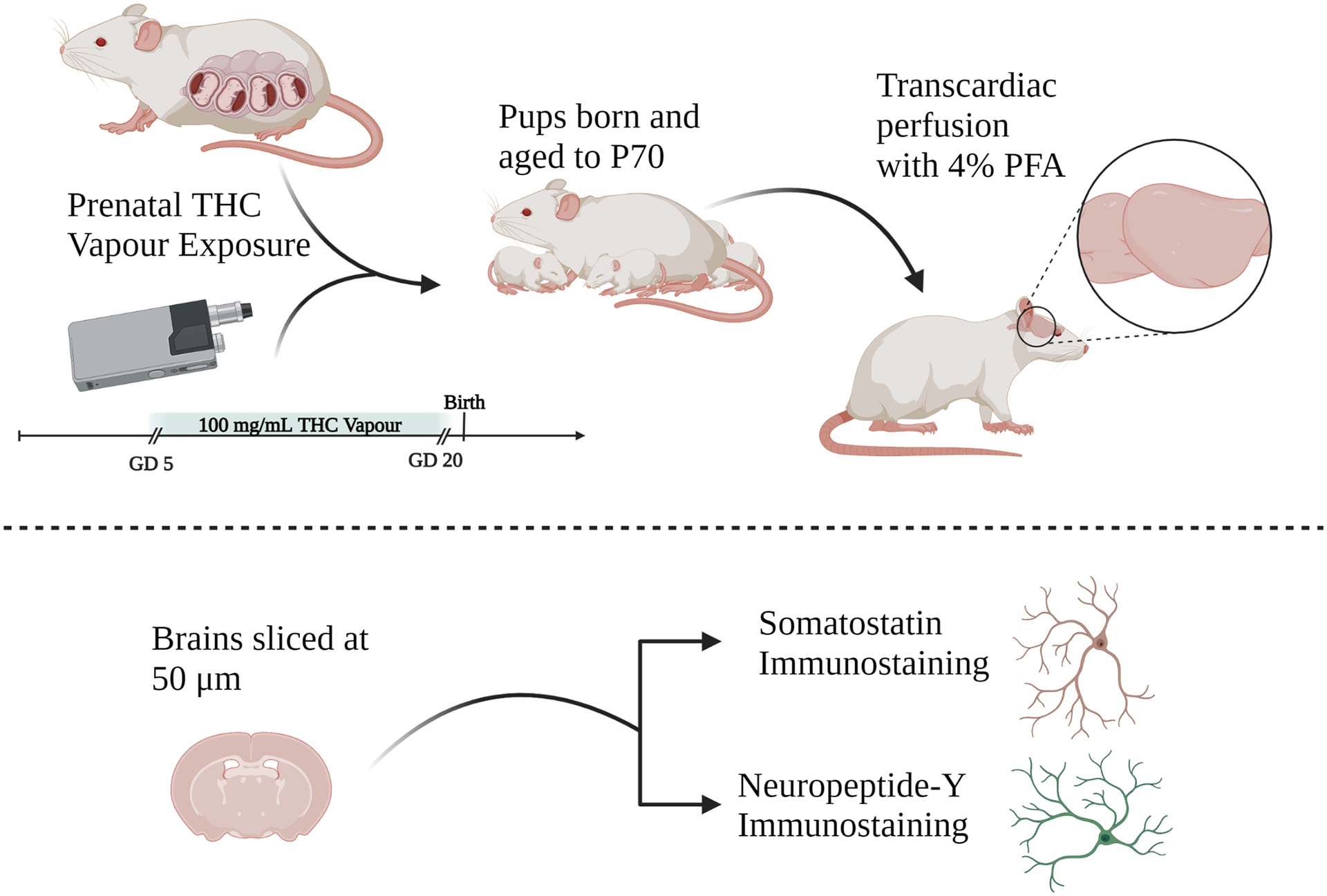
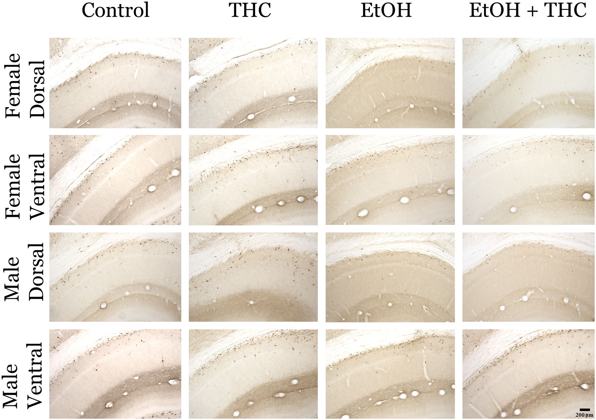
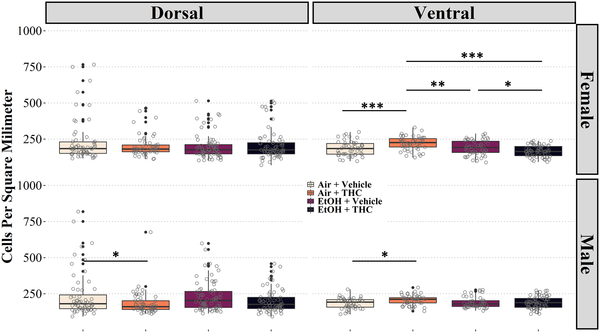
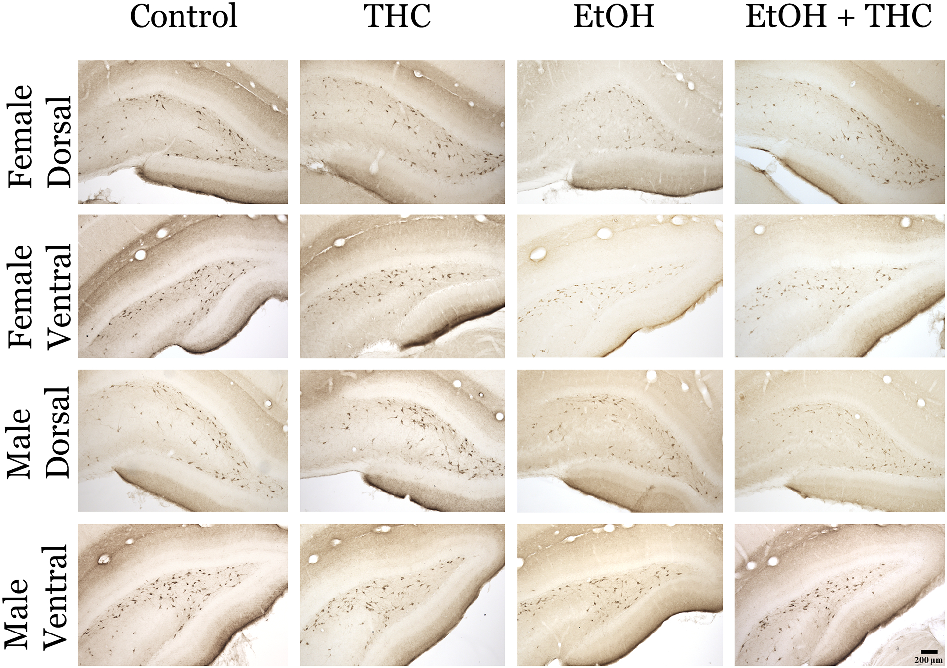
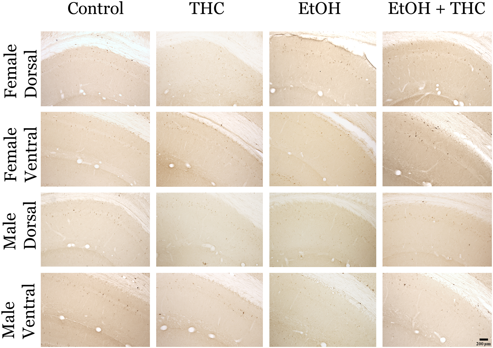
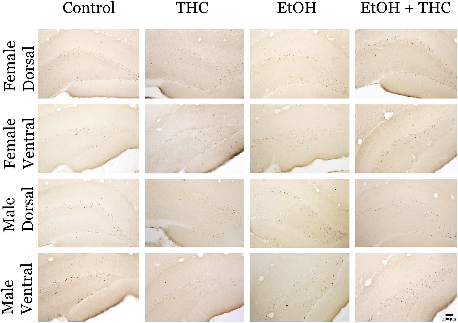
Methods: A 2 (Ethanol, Air) × 2 (tetrahydrocannabinol [THC], Vehicle) design was used to expose pregnant Sprague-Dawley rats to either ethanol or air, in addition to either THC or the inhalant vehicle solution, during gestational days 5-20. Immunohistochemistry for somatostatin- and NPY-positive interneurons was performed in 50 μm tissue sections obtained at postnatal day 70.

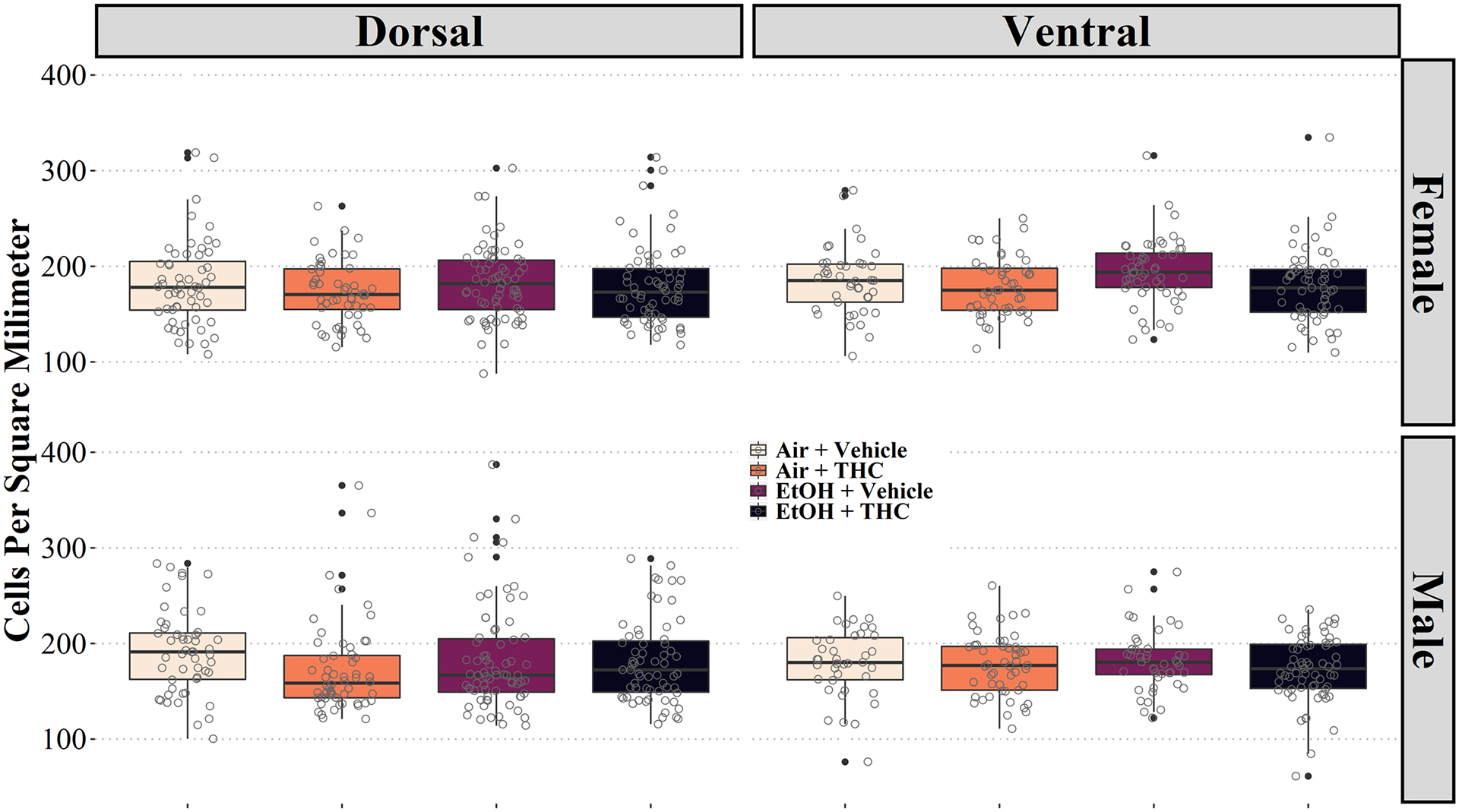
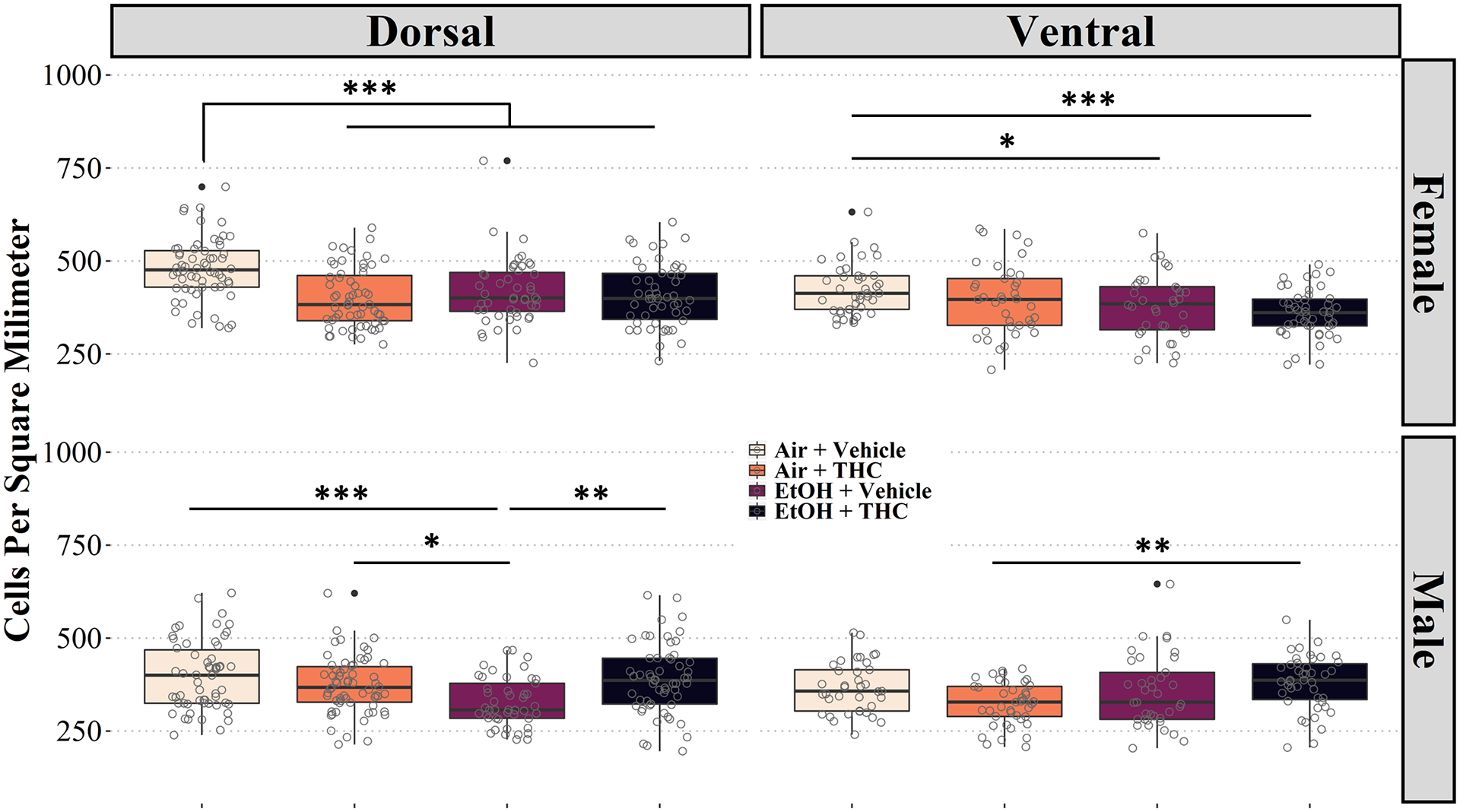
Results: Exposure to THC in utero had region-specific and sex-specific effects on the density of somatostatin-positive interneurons in the adult rat hippocampus. A female-specific decrease in NPY interneuron cell density was observed in the CA1 region following THC exposure. Combined exposure to alcohol and THC reduced NPY neurons selectively in the ventral dentate gyrus hippocampal subfield. However, overall, co-exposure to alcohol and cannabis had neither additive nor synergistic effects on interneuron populations in other areas of the hippocampus.

Conclusions: These results illustrate how alcohol and cannabis exposure in utero may affect hippocampal function by altering inhibitory processes in a sex-specific manner.

Keywords: alcohol; cannabis; dentate gyrus; hippocampus; interneuron.


Figures

Figure 1:
Figure 1:. Schematic showing the progression of techniques employed for these experiments.
Pregnant rats were exposed to 95% EtOH or Air for 3 consecutive hours at an airflow of 10 L/min. Afterwards, all rats were either exposed to THC or propylene glycol vehicle at an airflow rate of 2mL/min. Blood samples were taken on GDs 5, 10, 15, and 20. Between PD60–70, the adult offspring were deeply anesthetized with lethal ketamine (67 mg/mL) and xylazine (6.7 mg/mL) and were perfused through the heart. Brains were then collected and post-fixed in 4% paraformaldehyde for 24 hours before being sliced at 50 μm for histological analysis.
Figure 2:
Figure 2:. Somatostatin Positive Interneurons in Dorsal and Ventral CA1 Subfield of the Hippocampus.
Photomicrographs of representative images of somatostatin interneurons in the CA1 of male and female rats in each exposure group. Images were taken at 10x (0.30 NA) on an Olympus BX61 microscope.
Figure 3:
Figure 3:. Prenatal alcohol and cannabis combined does not produce synergistic changes in somatostain positive neurons in the CA1 subfield of the hippocampus.
Box and whisker plots showing the calculated density of somatostatin-positive cells in rats exposed to air, THC, EtOH, and EtOH + THC from GD5–20 in the dorsal and ventral CA1. There was a significant increase in the density of SOM interneurons in the ventral CA1 of male and female animals (p = 0.05 and p < 0.001) in the THC exposed group relative to control; however, in females the THC group was also significantly higher than both the ethanol (EtOH) group (p = 0.005) as well as the combined exposure group (p < 0.001). The EtOH group was also significantly increased relative to the combined group in the ventral CA1 of females (p = 0.035). Data collected as profile counts of a DAB stained interneurons from a 1 in 6 series of tissue sections. Densities were calculated from area measurements of taken from each section..
Figure 4:
Figure 4:. Somatostatin Positive Interneurons in Dorsal and Ventral DG Subfield of the DG subfield of the Hippocampus.
Photomicrographs of representative images of somatostatin interneurons in the DG of male and female rats in each exposure group. Images were taken at 10x (0.30 NA) on an Olympus BX61 microscope.
Figure 5:
Figure 5:. No significant changes in somatostatin interneurons in the DG following prenatal THC and/or ethanol exposure.
Calculated density of somatostatin-positive cells in rats exposed to air, THC, EtOH, and EtOH + THC from GD5–20 in the dentate gyrus. Data collected as profile counts of a DAB stained interneurons from a 1 in 6 series of tissue sections. Densities were calculated from area measurements of taken from each section.
Figure 6:
Figure 6:. Neuropeptide Y Positive Interneurons in Dorsal and Ventral CA1 Subfield of the Hippocampus.
Photomicrographs of representative images of neuropeptideY interneurons in the CA1 of male and female rats in each exposure group. Images were taken at 10x (0.30 NA) on an Olympus BX61 microscope.
Figure 7:
Figure 7:. Prenatal exposure to THC and/or ethanol causes sex-specific changes in NPY-positive interneurons.
Graphs depict the calculated density of NPY-positive cells in rats (post-natal day 70) exposed to air, THC, EtOH, and EtOH + THC from GD5–20 in the dorsal and ventral CA1. In the CA1, females showed a significant decrease in the density of NPY interneurons with THC in both dorsal (p = 0.011) and ventral (p = 0.20) regions. In the dorsal region, NPY interneuron density was also decreased in females exposed to EtOH only (p = 0.009). Ventrally, the decrease in the THC group was also significantly different than the combined exposure group (p = 0.005). Data collected as profile counts of a DAB stained interneurons from a 1 in 6 series of tissue sections. Densities were calculated from area measurements of taken from each section.
Figure 8:
Figure 8:. Neuropeptide Y Positive Interneurons in Dorsal and Ventral CA1 Subfield of the Hippocampus.
Photomicrographs of representative images of neuropeptideY interneurons in the DG of male and female rats in each exposure group. Images were taken at 10x (0.30 NA) on an Olympus BX61 microscope.
Figure 9: :
Figure 9: :. Neuropeptide Y Positive Interneurons are reduced in Dorsal and Ventral DG Subfield of the Hippocampus in a sex-specific manner.
Graphs depict the calculated density of NPY-positive cells in rats exposed to air, THC, EtOH, and EtOH + THC from GD5–20 in the dentate gyrus. In the dorsal dentate gyrus of males, the EtOH group was significantly decreased compared with all other groups (control (p < 0.001), THC (p = 0.030), and combined exposure (p = 0.002)). The only interaction in the ventral dentate gyrus of males was a higher density in the combined exposure group relative to the THC group (p = 0.004). In the dorsal dentate gyrus of females, the density of NPY interneurons was decreased in all three drug-exposed groups relative to control (p < 0.001 in all cases). Ventrally, there was a decrease in density between controls and EtOH (p = 0.049) as well as controls and combined exposure (p < 0.001). Data collected as profile counts of a DAB stained interneurons from a 1 in 6 series of tissue sections. Densities were calculated from area measurements of taken from each section..

Similar articles

Cited by

References

    1. Abbas AI, Sundiang MJM, Henoch B, Morton MP, Bolkan SS, Park AJ, Harris AZ, Kellendonk C, Gordon JA, 2018. Somatostatin Interneurons Facilitate Hippocampal-Prefrontal Synchrony and Prefrontal Spatial Encoding. Neuron 100, 926–939.e3. 10.1016/j.neuron.2018.09.029 - DOI - PMC - PubMed
    1. Adrian TE, Allen JM, Bloom SR, Ghatei MA, Rossor MN, Roberts GW, Crow TJ, Tatemoto K, Polak JM, 1983. Neuropeptide Y distribution in human brain. Nature 306, 584–586. 10.1038/306584a0 - DOI - PubMed
    1. Alviña K, Jodeiri Farshbaf M, Mondal AK, 2021. Long term effects of stress on hippocampal function: Emphasis on early life stress paradigms and potential involvement of neuropeptide Y. Journal of Neuroscience Research 99, 57–66. 10.1002/jnr.24614 - DOI - PubMed
    1. Andersen P, Morris R, Amaral D, Bliss T, O’Keefe J, 2007. The Hippocampus Book. Oxford University Press, Oxford.
    1. Andrade JP, Fernando PM, Madeira MD, Paula‐Barbosa MM, Cadete‐Leite A, Zimmer J, 1992. Effects of chronic alcohol consumption and withdrawal on the somatostatin-immunoreactive neurons of the rat hippocampal dentate hilus. Hippocampus 2, 65–71. 10.1002/HIPO.450020109 - DOI - PubMed

LinkOut - more resources