. 2024 May 24;15(1):4444.
doi: 10.1038/s41467-024-48653-y.

Deleting the mitochondrial respiration negative regulator MCJ enhances the efficacy of CD8+ T cell adoptive therapies in pre-clinical studies

Affiliations

Deleting the mitochondrial respiration negative regulator MCJ enhances the efficacy of CD8+ T cell adoptive therapies in pre-clinical studies

Meng-Han Wu et al. Nat Commun. .

Abstract

Mitochondrial respiration is essential for the survival and function of T cells used in adoptive cellular therapies. However, strategies that specifically enhance mitochondrial respiration to promote T cell function remain limited. Here, we investigate methylation-controlled J protein (MCJ), an endogenous negative regulator of mitochondrial complex I expressed in CD8 cells, as a target for improving the efficacy of adoptive T cell therapies. We demonstrate that MCJ inhibits mitochondrial respiration in murine CD8+ CAR-T cells and that deletion of MCJ increases their in vitro and in vivo efficacy against murine B cell leukaemia. Similarly, MCJ deletion in ovalbumin (OVA)-specific CD8+ T cells also increases their efficacy against established OVA-expressing melanoma tumors in vivo. Furthermore, we show for the first time that MCJ is expressed in human CD8 cells and that the level of MCJ expression correlates with the functional activity of CD8+ CAR-T cells. Silencing MCJ expression in human CD8 CAR-T cells increases their mitochondrial metabolism and enhances their anti-tumor activity. Thus, targeting MCJ may represent a potential therapeutic strategy to increase mitochondrial metabolism and improve the efficacy of adoptive T cell therapies.


Conflict of interest statement

M.R. declares to be a member of the scientific advisory board and co-founder of Mitotherapeutix, and is co-inventor in patents held by the University of Vermont. M.R., M.E.K., M.W. and M.C.Y. declare the filing of a patent that discloses findings described in this manuscript. R.A.F. is an advisor to Glaxo Smith Kline, Celsius, EvolveImmune, Ventus Therapeutics, and is the recipient of a grant from Genetech/Roche. The remaining authors declare no competing interests.

Figures
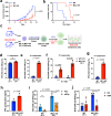
Fig. 1
Fig. 1. Loss of MCJ in TCR-antigen specific CD8 cells enhances their anti-tumor killing activity in vitro.
a, b B16-OVA melanoma cells (4 × 105/mouse) were subcutaneously (s.c.) injected on the flank of WT or MCJ KO host mice. Tumor volume over time (a) and survival (b) were followed (n = 5 biologically independent animals). cf WT or MCJ KO OT-I CD8 cells were activated for 2 days with anti-CD3/anti-CD28 Abs, washed, and then expanded with IL-2 (40 IU/ml) for 2 days (1st expansion) and expanded again with fresh medium and IL-2 (2nd expansion) (c) (created using BioRender.com). After the 1st expansion with IL-2, WT and MCJ KO OT-I cells were co-cultured with B16-OVA cells for killing assay at an E:T = 10, and live B16-OVA cells were counted after 24 h (n = 3 biologically independent cells) (d). After the 2nd expansion with IL-2, WT and MCJ KO OT-I cells were co-cultured with B16-OVA cells for killing assay at an E:T = 10 and live B16-OVA cells were counted after 24 h (n = 3 biologically independent cells) (e), or an E:T = 1 and E:T = 0.1 and live B16-OVA cells were counted after 46 h co-culture (n = 3 biologically independent cells) (f). g WT and MCJ KO OT-I cells were activated as in (c) and after the 3rd expansion with IL-2 were co-cultured with B16-OVA cells at an E:T = 5. Live B16-OVA cells were counted after 24 h co-culture (n = 3 biologically independent cells). h ELISA of IFNγ levels in supernatant of co-cultures of B16-OVA cells with either WT or MCJ KO CD8 OT-I cells after the 2nd expansion (n = 4 biologically independent samples). i WT and MCJ KO OT-I cells were activated, expanded with IL-2 for two expansions and co-cultured with B16-OVA cells at an E:T = 10, with or without an anti-IFNγ blocking Ab. Live B16-OVA cells were counted after 24 h (n = 3 biologically independent cells). j WT and MCJ KO OT-I cells were activated and expanded as in (i), pretreated with CMA or vehicle for 2 h, washed, and co-cultured with B16-OVA cells (E:T = 2). Live B16-OVA cells were counted after 24 h (n = 4 biologically independent cells). p was determined by mixed-effect analysis (a), Mantel-Cox test (b), two-sided unpaired t test (dh), and 2-way ANOVA multiple comparisons (i, j). Mean ± SD is shown for (a, dj).
Fig. 2
Fig. 2. Superior efficacy of MCJ-deficient TCR-antigen specific CD8 cells for the treatment of melanoma in vivo.
a B16-OVA tumor cells (4 × 105/mouse) were s.c. injected on the flank to WT mice (n = 7 biologically independent animals per group). WT and MCJ KO OT-I CD8 cells were activated with anti-CD3/anti-CD28 Abs as in Fig. 1, expanded with IL-2 for two expansions, and (5 × 105 cells/mouse) i.v. administered to the B16-OVA tumor-bearing mice 10 days post-implantation when the tumors were palpable. Tumor size was followed for 8 days (when a large fraction of mice needed to be euthanized based on tumor size), and it is presented as fold-increase relative to the size of the tumor at the time of the CD8 cell adoptive transfer. b Immunostaining for CD8 (red) and DAPI as nuclear marker (blue) of histological sections from B16-OVA tumors harvested 8 days after the adoptive transfer of WT or MCJ KO OT-I CD8 cells, as in (a). ce WT and MCJ KO OT-I CD8 cells were activated and expanded as in (a), and injected into WT mice (n = 5 biologically independent animals per group) bearing B16-OVA melanoma tumors. Tumors were harvested 10 days after CD8 cell adoptive transfer and tumor cell homogenate was examined by flow cytometry for the presence of total CD8 cells (c) or OT-I CD8 cells (d) within the leukocyte (CD45+) population, as well as the expression of CD44 and PD1 on OT-I CD8 cells within the OT-I CD8 cells (e). f WT and MCJ KO OT-I CD8 cells were activated and expanded as in (a), and injected into WT mice (n = 4 biologically independent animals per group) bearing B16-OVA tumors. 7 days after CD8 cell adoptive transfer tumors were harvested to isolate tumor infiltrating CD8 cells. Pooled infiltrated CD8 cells were and activated ex vivo with anti-CD3/anti-CD28 Abs for 24 h and IFNγ levels in supernatant was examined by ELISA (n = 3 replicate wells per group). p was determined by 2-way ANOVA analysis (a) and two-sided unpaired t test (cf). Mean ± SD is shown for (a, cf).
Fig. 3
Fig. 3. MCJ-deficient CD8 CAR-T cells are superior in perforin-mediated killing of leukemia B cells in vitro.
a WT or MCJ KO CD8 cells were activated with anti-CD3/anti-CD28 Abs for 1 day, retrovirally transduced with CD19-BBz CAR, expanded with IL-2 (60 IU/ml) for 2 days (1st expansion), and co-cultured with E2a leukemia cells (E:T = 0.5) for killing assay. Live E2a cells were examined by flow cytometry after 15 h (n = 3 biologically independent cells). b WT or MCJ KO CD8 cells were activated and transduced with CD19-BBz CAR as in (a). After three expansions with IL-2, CD8 CAR-T cells were co-cultured with E2a leukemia cells (E:T = 0.5) for killing assay (n = 3 biologically independent cells). c WT or MCJ KO CD8 cells were activated and transduced with CD19-BBz CAR. After the 3rd expansion with IL-2, cells were washed, and incubated in medium along for 24 h. CAR-T cells were then co-cultured with E2a leukemia cells at the given E:T ratio for a 5 h killing assay (n = 3 biologically independent cells). d IFNγ production during the killing assay with WT and MCJ KO CD8 CAR-T cells generated as in (b) and E2a leukemia cells (E:T = 0.5), as determined by ELISA (n = 3 biologically independent cells). (e) IFNγ production during the killing assay with WT and MCJ KO CD8 CAR-T cells generated as in (c) and E2a leukemia cells (E:T = 0.3) (n = 3 biologically independent cells). f WT or MCJ KO CD8 cells were activated and transduced with CD19-BBz CAR as in (a). After the 3rd expansion with IL-2, cells were washed, and incubated in medium alone for 24 h. CAR-T cells were then co-cultured with E2a leukemia cells (E:T = 0.3) in the presence or absence of an anti-IFNγ blocking Ab for a killing assay (n = 3 biologically independent cells). g CD107a expression on 3rd-expansion WT and MCJ KO CD8 CAR-T cells after 4 h of co-culture with E2a cells, as determined by flow cytometry. % of CD107a+ cells is shown. h After the 3rd expansion with IL-2 followed by 24 h resting in medium, WT and MCJ KO CD8 CAR-T cells were co-cultured with E2a cells (E:T = 0.3) in the presence or the absence of EGTA (3 mM)/MgCl2 (2 mM). Live E2a cells were counted after 5 h (n = 3 biologically independent cells). i After the 3rd expansion with IL-2 followed by 24 h resting in medium, WT and MCJ KO CD8 CAR-T cells were pre-treated with CMA (100 nM) or vehicle for 2 h, washed and co-cultured with E2a cells (E:T = 0.4) for 5 h. Live E2a cells were counted (n = 3 biologically independent cells). p  was determined by two-sided unpaired t test (ae) or 2-way ANOVA (f, h, i). Mean ± SD is shown for all figures with bar graph.
Fig. 4
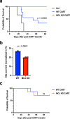
Fig. 4. Lack of MCJ in CD8 CAR-T cells improves their efficacy against the leukemia B cells in vivo.
a E2a cells (106/mouse) were injected i.v. into WT host mice (n = 9 biologically independent animals per group). 3 days later, mice were sublethally irradiated (500 cGy), and the next day they received an i.v. administration of PBS alone as vehicle (Ctrl), WT CD19-BBz CD8 CAR-T cells or MCJ KO CD19-BBz CD8 CAR-T cells (105 CAR-T cells/mouse) that were expanded with IL-2 for 3 expansions. Survival of the mice post-treatment with CAR-T cells was recorded over time. Kaplan-Meier survival analysis is shown. b WT host mice were administered with E2a cells, irradiated and treated with WT or MCJ KO CD19-BBz CD8 CAR-T cells (106 CAR-T cells/mouse) as described in (a). 21 days after the treatment with CAR-T cells, bone marrow was harvested to isolate CD8 cells that were then used for an ex vivo killing assay with E2a cells as targets (E:T = 0.05, based on the percentage of the CAR + CD8 cells). Same number of CAR + CD8 cells were co-cultured with E2a cells. Live E2a cells were counted after 20 h co-culture (n = 3 replicate wells per group). c Kaplan–Meier survival analysis of leukemia-bearing mice treated as in (a) with WT or MCJ KO CD19-BBz CD8 CAR-T cells after the 1st expansion with IL-2 (n = 7 biologically independent animals per group). p  was determined by log-rank (Mantel–Cox) test (a, c) or two-sided unpaired t test (b). Mean ± SD is shown for (b).
Fig. 5
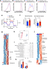
Fig. 5. MCJ deficiency enhances the mitochondrial fitness of CD8 CAR-T cells.
ad WT or MCJ KO CD8 cells were activated with anti-CD3/anti-CD28 Abs, transduced with CD19-BBz CAR, and expanded with IL-2. After the 1st expansion (a and b) or the 3rd expansion (c and d) with IL-2, cells were stained with TMRE (a and c) or Mitotracker (b and d) and analyzed by flow cytometry. Numbers in parenthesis show the geometric mean fluorescence intensity. e, f After three expansions with IL-2, WT and MCJ KO CAR-T cells (as in (a)) were purified and used for Seahorse MitoStress assay. OCR was measured at baseline and in response to oligomycin (Oligo), FCCP and rotenone with antimycin (R/A) (n = 16 replicate wells per group) (e). Maximal respiration and spare respiratory capacity (f) were determined. g, h Mass spectrometry-based metabolomics analysis of purified WT and MCJ KO CD8 CAR-T cells after three expansions with IL-2 (n = 4 replicate cells per group). Hierarchical clustering analysis of the relative abundance of top 50 distinct metabolites shown in heat map from two groups. Metabolites of interests are highlighted (g). PCA of two groups (h). i WT or MCJ KO CD8 cells were activated and transduced to generate CD19-BBz CAR-T cells as in (a). After 3 expansions with IL-2, WT and MCJ KO CAR-T cells are purified and used for RNAseq. The top 10 distinctly differential pathways in GSEA HALLMARK pathways are shown as water plot. j Mass spectrometry-based metabolomics analysis of purified WT and MCJ KO CD8 CAR-T cells after three expansions with IL-2 followed by 48 h in cytokine-free medium (n = 3 replicate cells per group). Hierarchical clustering analysis of the relative abundance of top 40 distinct metabolites between two groups shown in heat map. Metabolites of interests are highlighted. k After 3 expansions with IL-2 followed by 24 h in cytokine-free medium, WT and MCJ KO CD8 CAR-T cells were pretreated with oligomycin (5 μM) or DMSO (Veh) for 3 h, washed and co-cultured with E2a cells for 4 h for killing assay (n = 3 biologically independent cells). p  was determined by two-way ANOVA (e, k), two-sided unpaired t test (f, g, j). i p-adj was calculated using Benjamini–Hochberg procedure using 10,000 permutations. Mean ± SD is shown (e, f, k).
Fig. 6
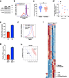
Fig. 6. MCJ regulates mitochondrial metabolism and effector function in human CD8 cells.
a MCJ expression by Western blot analysis using whole cell extracts from CD4 and CD8 cells freshly isolated from peripheral blood of a healthy volunteer. NDUFA9 subunit of Complex I was examined as a mitochondria content normalization control. b MMP by TMRE staining in combination with staining for CD4 and CD8 of freshly obtained PBMC by flow cytometry. The percentage of TMREhigh cells is shown in parenthesis. c Data from normalized MCJ/DnaJC15 mRNA levels (transcript per million, TPM) in human naive CD8 cells from healthy subjects (54 males and 35 females) were obtained from DICE database. d MCJ/DnaJC15 mRNA levels in freshly isolated CD8 cells from 6 male (M) and 5 female (F) healthy volunteers assayed by real-time RT-PCR. β2-microglobulin was used as housekeeping gene for normalization. e, f MCJ mRNA levels in freshly isolated CD8 cells from healthy donors H3 and H6 were examined by real-time RT-PCR using HPRT as housekeeping gene for normalization (n = 2 replicates for each donor) (e). CD8 cells from both H3 and H6 donors were activated with anti-human CD3 (10 μg/ml) and anti-human CD28 (2 μg/ml) Abs for 2 days, expanded with IL-2 (100 IU/ml) and after two expansions MMP was examined by TMRE staining. Numbers in parenthesis show the mean fluorescence intensity (f). gi CD8 cells were isolated from healthy donors D16 and D26 and MCJ mRNA levels were assayed by real time RT-PCR, using HPRT as housekeeping gene (n = 2 replicates for each donor) (g). CD8 cells from both donors were activated with anti-CD3/anti-CD28 Abs, transduced with a human CD19-BBz CAR, and expanded with IL-2. After three expansions, purified CAR-T cells were used for metabolomics analysis (n = 3 replicate cells per group). Partial least squares-discriminant analysis (PLS-DA) of D16 (MCJlow) and D26 (MCJhigh) CAR-T cells (h). Hierarchical clustering analysis of the relative abundance of top 45 distinct metabolites shown in heat map from D16 (MCJlow) and D26 (MCJhigh) CAR-T cells. Metabolites of interests are highlighted (i). p was determined by two-sided unpaired t test (c, i). Mean is shown for (c). Mean ± SD is shown for (e, g).
Fig. 7
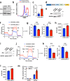
Fig. 7. MCJ deficiency improves human CD19-BBz CAR-T cell efficacy.
a CD8 cells isolated from the PBMC of a healthy volunteer were transfected by nucleofection with a control siRNA or siMCJ and activated with coated anti-CD3/anti-CD28 for 2 days. MCJ levels were analyzed by Western blot analysis. b CD8 cells from a healthy donor were transfected with siMCJ or control siRNA as in (a), activated for 2 days, expanded with IL-2 for 2 days. MMP of the cells was examined by TMRE staining. The percentage of TMREhigh cells is shown in parenthesis. c CD8 cells from a healthy donor were transfected and activated as in (a), washed, and incubated in medium alone for 48 h. IFNγ levels in the supernatant was determined by ELISA (n = 3 biologically independent samples). d Scheme (created using BioRender.com) showing the human CD19-BBz/shMCJ CAR construct containing (a) the scFv part of human CD19 (hCD19 scFv), (b) the transmembrane domain (TM) of human CD8 (hCD8 TM), (c) the cytoplasmic costimulatory domain of human 4-1BB (h41BB), (d) the cytoplasmic domain of human CD3z (hCD3z) and the shRNA for human MCJ cassette (including the U6 promoter). As a control we used the same construct but with a scramble shRNA. e CD8 cells were isolated from a healthy donor, activated with anti-CD3/anti-CD28 Abs for 2 days, transduced with the lentiviral CD19-BBz/shMCJ-1 CAR or CD19-BBz/shRNA CAR construct, and expanded with IL-2 (100 IU/ml) for 3 expansions. MCJ expression in purified CD19-BBz/shMCJ-1 or CD19-BBz/c-shRNA CAR-T cells after 3 expansions, by Western blot analysis. f After 3 expansions with IL-2, purified CD19-BBz/shMCJ-1 or CD19-BBz/c-shRNA CD8 CAR-T cells were used for Seahorse MitoStress assay (n = 10 replicate well per group). g CD8 cells isolated from two healthy donors (H16 and H19), were activated, transduced and expanded as (e). After the 3rd expansion with IL-2, CD19-BBz/shMCJ-1 or CD19-BBz/c-shRNA CAR-T cells were co-cultured with the human B cell leukemia Nalm6 cells (E:T = 0.125). After 20 h, live Nalm6 cells were counted (n = 3 biologically independent cells). h CD107a expression (MFI) on CD19-BBz/shMCJ or CD19-BBz/c-shRNA CAR-T cells after 4 h of co-culture with Nalm6 cells, as determined by flow cytometry (n = 3 biological independent cells). i Human CD8 cells were activated, transduced with lentiviral construct contained CD19-BBz/shMCJ-2 CAR or CD19-BBz/c-shRNA CAR and expanded with IL-2 for 3 expansions. MCJ expression in purified CAR-T cells, by Western blot analysis. j After 3 expansions with IL-2, purified human CD19-BBz/shMCJ-2 or CD19-BBz/c-shRNA CD8 CAR-T cells were used for Seahorse MitoStress assay (n = 10 replicate wells per group). k CD8 cells isolated from two healthy donors (H16 and H19), were activated, transduced and expanded as (h), and CD19-BBz/shMCJ-2 or CD19-BBz/c-shRNA CAR-T cells were co-cultured with the human B cell leukemia Nalm6 cells (E:T = 0.125). After 20 h, live Nalm6 cells were counted (n = 3 biologically independent cells). l CD107a expression (MFI) on CD19-BBz/shMCJ-2 or CD19-BBz/c-shRNA CAR-T cells after 4 h of co-culture with Nalm6 cells (n = 3 biologically independent cells). m IFNγ production of CD19-BBz/shMCJ-2 or CD19-BBz/c-shRNA CAR-T cells during co-culture with Nalm6 cells at E:T = 0.25, as determined by ELISA (n = 3 biologically independent samples). p  was determined by two-sided unpaired t test (c, g, h, k, l) and by two-way ANOVA (f, j, m). Mean ± SD are shown for (c, fh, jm).

References

    1. June CH, Riddell SR, Schumacher TN. Adoptive cellular therapy: a race to the finish line. Sci. Transl. Med. 2015;7:280ps287. doi: 10.1126/scitranslmed.aaa3643. - DOI - PubMed
    1. Rohaan MW, Wilgenhof S, Haanen J. Adoptive cellular therapies: the current landscape. Virchows Arch. 2019;474:449–461. doi: 10.1007/s00428-018-2484-0. - DOI - PMC - PubMed
    1. Sterner RC, Sterner RM. CAR-T cell therapy: current limitations and potential strategies. Blood Cancer J. 2021;11:69. doi: 10.1038/s41408-021-00459-7. - DOI - PMC - PubMed
    1. Mitra A, et al. From bench to bedside: the history and progress of CAR T cell therapy. Front Immunol. 2023;14:1188049. doi: 10.3389/fimmu.2023.1188049. - DOI - PMC - PubMed
    1. Davila ML, et al. Efficacy and toxicity management of 19-28z CAR T cell therapy in B cell acute lymphoblastic leukemia. Sci. Transl. Med. 2014;6:224ra225. doi: 10.1126/scitranslmed.3008226. - DOI - PMC - PubMed