. 2024 May 24;14(1):11915.
doi: 10.1038/s41598-024-62106-y.

Choosing a camera and optimizing system parameters for speckle contrast optical spectroscopy

Affiliations

Choosing a camera and optimizing system parameters for speckle contrast optical spectroscopy

Tom Y Cheng et al. Sci Rep. .

Abstract

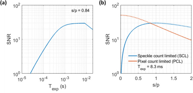
Speckle contrast optical spectroscopy (SCOS) is an emerging camera-based technique that can measure human cerebral blood flow (CBF) with high signal-to-noise ratio (SNR). At low photon flux levels typically encountered in human CBF measurements, camera noise and nonidealities could significantly impact SCOS measurement SNR and accuracy. Thus, a guide for characterizing, selecting, and optimizing a camera for SCOS measurements is crucial for the development of next-generation optical devices for monitoring human CBF and brain function. Here, we provide such a guide and illustrate it by evaluating three commercially available complementary metal-oxide-semiconductor cameras, considering a variety of factors including linearity, read noise, and quantization distortion. We show that some cameras that are well-suited for general intensity imaging could be challenged in accurately quantifying spatial contrast for SCOS. We then determine the optimal operating parameters for the preferred camera among the three and demonstrate measurement of human CBF with this selected low-cost camera. This work establishes a guideline for characterizing and selecting cameras as well as for determining optimal parameters for SCOS systems.


Conflict of interest statement

The authors declare no competing interests.

Figures

Figure 1
Figure 1
Characterization of camera gain and nonlinearity-induced K2 error for three CMOS cameras. (a) Schematic of the setup for measuring the cameras’ photon transfer curves. (bd) Estimation of camera gain and linearity error (LE) for each camera’s photon transfer curve. Here σ2 is the variance of the intensity and I is the average intensity across all the pixels, in units of the camera’s digital number (DN) output. Blue circles are the experimental data and red dashed lines are the linear regression lines. Error bars represent the temporal standard deviation of the variance of each difference image obtained. The linear regression is performed on data within a limited I range as indicated by the shaded region, where the shot noise is greater than twice the read noise and I is less than 70% of the camera’s saturation capacity. The estimated camera gain is the slope of the regression line. The LE is calculated as the mean magnitude of the relative deviation of the measured variance from the regression line within the fitting range. (eg) Estimation of the absolute nonlinearity-induced error in K2, |ΔK2|, within the shaded region indicated in (bd). Error bars represent the temporal standard deviation of the variance of each difference image divided by I2. HA: Hamamatsu Orca Fusion BT C15440-20UP; BAa: Basler a2A1920-160umPRO; BAd: Basler daA1280-54um.
Figure 2
Figure 2
Dark offset and read noise variance distributions for the three cameras. (a) Normalized mean dark offset Idark distributions across pixels for the three cameras in units of DN. Here N is the number of pixels at a particular dark offset, and it is normalized by its maximum value Nmax. The mean dark offsets of the cameras are HA = 100.5 DN, BAa = 49.4 DN, and BAd = 9.7 DN. (b) Normalized read noise variance σr2 distributions across pixels for the three cameras in units of DN2. The RMS read noise values of the cameras are HA = 5.99 DN, BAa = 1.16 DN, and BAd = 0.48 DN. (c) Normalized read noise variance σr2 distributions for the three cameras in units of e−2. The RMS read noise values of the cameras are HA = 1.28 e, BAa = 1.97 e, and BAd = 10.33 e.
Figure 3
Figure 3
Impact of quantization distortion on SCOS measurement accuracy. (a) Maximum quantization-distortion-induced error in measured mean intensity I as a function of the true variance σtrue2. (b) Maximum quantization-distortion-induced error in measured variance σ2 as a function of the true variance σtrue2. The error in σ2 asymptotically approaches 1/12 (red dashed line) with increasing σtrue2. (c) Corresponding error in K2 given the errors in I (a) and σ2 (b) as a function of σtrue2, after subtracting an assumed quantization-induced bias of 1/12 from the signal’s variance.
Figure 4
Figure 4
Dark offset and read noise variability of the BAa camera. (a) Time course of pixel-averaged mean dark offset Idark and read noise variance σr2. Dark measurements were performed every 10 min, and prior to the first measurement the camera was powered on but not acquiring images. Error bars represent the temporal standard deviation of the mean of each image (top) and the variance of each difference image (bottom). (b) Change in the Idark distribution across pixels between the two time points indicated. (c) Change in the σr2 distribution across pixels between the same two time points.
Figure 5
Figure 5
SCOS SNR dependence on exposure time and s/p ratio for the BAa camera. (a) SNR versus Texp for s/p = 0.84. (b) SNR versus s/p at Texp = 8.3 ms for speckle-count-limited and pixel-count-limited cases. When the SCOS system is speckle-count-limited, the total number of speckles/fiber modes is fixed at M = 3.3×10⁶, estimated from the area of the fiber output image and the s/p ratio obtained experimentally. In the speckle-count-limited case we assume that the camera has enough pixels to image all the speckles. When the system is pixel-count-limited, the total number of pixels is fixed at 1936 × 1216 pixels for the BAa camera, and we assume all pixels are filled with speckles. The SNR vs. s/p of our fiber-based SCOS system follows the solid red and blue lines. BAa camera parameter values of 1.97 e RMS read noise, 16% quantum efficiency (QE) at 852 nm wavelength, 120 Hz maximum frame rate (fmax) at 10-bit depth, and 1936 × 1216 pixels were used. An average photon flux per speckle of 22,619 s-1 (as derived in the Methods) and a measurement rate of 10 Hz were used.
Figure 6
Figure 6
Impact of varying the laser pulsing factor on SCOS SNR with the BAa camera. (ab) SNR as a function of s/p and Texp at (a) PF = 1 and (b) PF = 10 for the speckle-count-limited case. The total number of speckles/fiber modes is fixed at M = 3.3×10⁶ as done in Fig. 5, and we assume the camera has enough pixels to image all the speckles. (de) SNR as a function of s/p and Texp at (d) PF = 1 and (e) PF = 10 for the pixel-count-limited case. The total number of pixels is fixed at 1936 × 1216 pixels, and we assume all pixels are filled with speckles. The optimal SNR and corresponding K2 terms are plotted as a function of laser PF for both the (c) speckle-count-limited case and (f) pixel-count-limited case. In both cases, the SNR plateaus as the blood-flow-induced speckle contrast Kf2 dominates and the fundamental noise σKf2 becomes the dominant noise source. BAa camera parameter values of 1.97 e RMS read noise, 16% quantum efficiency (QE) at 852 nm wavelength, 120 Hz maximum frame rate (fmax) at 10 bit depth, and 1936 × 1216 pixels were used. An average photon flux per speckle of 22,619 s-1 (as derived in the Methods) and a measurement rate of 10 Hz were used.
Figure 7
Figure 7
Cardiac and mental subtraction measurements using the BAa camera with optimal operating parameters. (a) Diagram of the SCOS measurement setup. Brain and skull image is obtained from Wikimedia Commons. All the other elements of the image are generated by the authors using Microsoft PowerPoint. (bc) Recovery of the pulsatile waveform in (b) mean intensity I and (c) BFi = 1/Kf2 using SCOS. The three peaks (P1, P2, P3) and dicrotic notch are clearly visible and labeled in the BFi waveform. (de) Significant brain activation and recovery to baseline can be seen in both the (d) ΔOD=log10(I0/I(t)) (p = 2.04 × 10–7) and (e) relative change in BFi (p = 1.06 × 10–5). Shown are the average ΔOD and Δ BFi waveforms from fifteen trials. The green rectangular shaded region represents the time duration of a trial. The red shaded error region represents the standard error across the 15 trials.

References

    1. Bandera E, et al. Cerebral blood flow threshold of ischemic penumbra and infarct core in acute ischemic stroke. Stroke. 2006;37:1334–1339. doi: 10.1161/01.STR.0000217418.29609.22. - DOI - PubMed
    1. Leigh R, Knutsson L, Zhou J, van Zijl PC. Imaging the physiological evolution of the ischemic penumbra in acute ischemic stroke. J. Cereb. Blood Flow Metab. 2018;38:1500–1516. doi: 10.1177/0271678X17700913. - DOI - PMC - PubMed
    1. Aracki-Trenkic A, et al. ASL perfusion in acute ischemic stroke: The value of CBF in outcome prediction. Clin. Neurol. Neurosurg. 2020;194:105908. doi: 10.1016/j.clineuro.2020.105908. - DOI - PubMed
    1. Bouma GJ, Muizelaar JP. Cerebral blood flow, cerebral blood volume, and cerebrovascular reactivity after severe head injury. J. Neurotrauma. 1992;9(Suppl 1):S333–348. - PubMed
    1. Kelly DF, et al. Cerebral blood flow as a predictor of outcome following traumatic brain injury. J. Neurosurg. 1997;86:633–641. doi: 10.3171/jns.1997.86.4.0633. - DOI - PubMed