. 2024 May 24;14(1):11909.
doi: 10.1038/s41598-024-62871-w.

Fluorescence tracking demonstrates T cell recirculation is transiently impaired by radiation therapy to the tumor

Affiliations

Fluorescence tracking demonstrates T cell recirculation is transiently impaired by radiation therapy to the tumor

Gwen Kramer et al. Sci Rep. .

Abstract

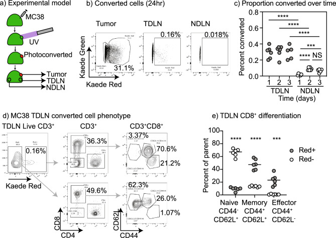
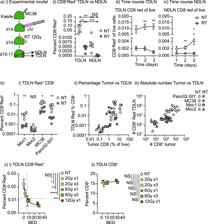
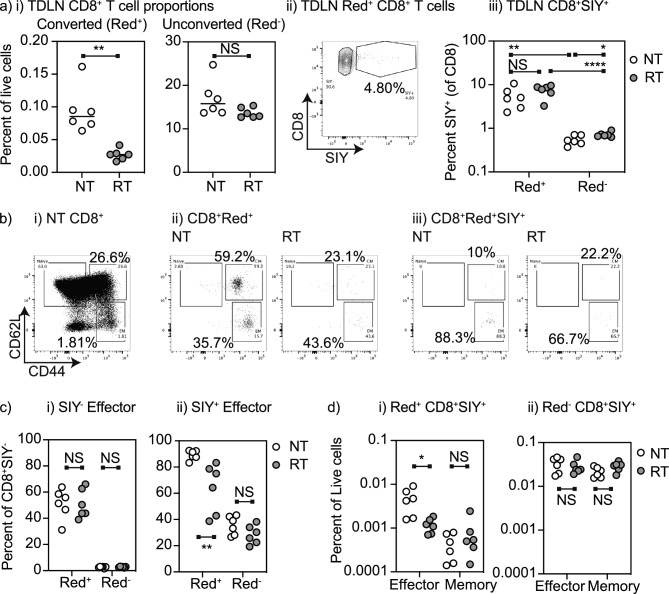
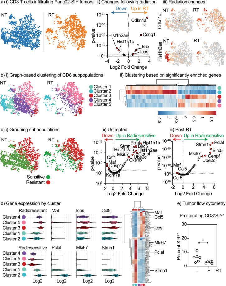
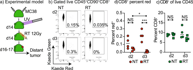
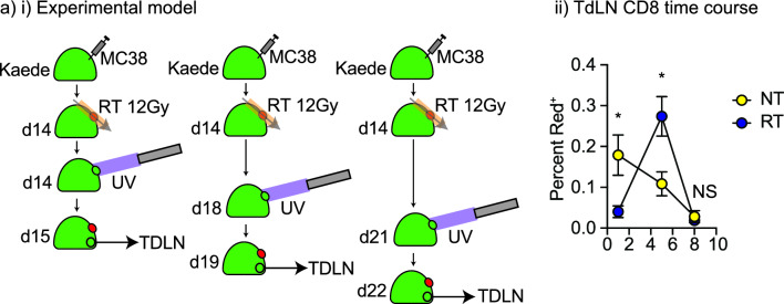
T cells recirculate through tissues and lymphatic organs to scan for their cognate antigen. Radiation therapy provides site-specific cytotoxicity to kill cancer cells but also has the potential to eliminate the tumor-specific T cells in field. To dynamically study the effect of radiation on CD8 T cell recirculation, we used the Kaede mouse model to photoconvert tumor-infiltrating cells and monitor their movement out of the field of radiation. We demonstrate that radiation results in loss of CD8 T cell recirculation from the tumor to the lymph node and to distant sites. Using scRNASeq, we see decreased proliferating CD8 T cells in the tumor following radiation therapy resulting in a proportional enrichment in exhausted phenotypes. By contrast, 5 days following radiation increased recirculation of T cells from the tumor to the tumor draining lymph node corresponds with increased immunosurveillance of the treated tumor. These data demonstrate that tumor radiation therapy transiently impairs systemic T cell recirculation from the treatment site to the draining lymph node and distant untreated tumors. This may inform timing therapies to improve systemic T cell-mediated tumor immunity.


Conflict of interest statement

The research was funded by NCI R01CA182311, NCI R01CA244142, and R01CA208644, and by the Providence Foundation. MJG receives research funding from Bristol Myers Squibb. MRC receives consulting fees from Roche. All other authors have no conflicts to declare. Research funding is not directly related to the topic of this manuscript. Funders had no role in the representation of the data or the preparation of the manuscript.

Figures

Figure 1
Figure 1
Identification of tumor origin recirculating CD8 T cells in lymph nodes. (a) Kaede mice were implanted with MC38 tumors and at d14 tumors were selectively photoconverted with UV light. Tumors, TdLN and NdLN were harvested over time. (b) Identification of photoconverted Kaede Red cells in the tumor, TdLN, and NdLN 24 h following photoconversion of the tumor. (c) Percent of TdLN and NdLN cells that are photoconverted over time. (d) Phenotype of photoconverted CD3+ cells in the TdLN at 24 h. (e) Quantification of the proportion of Photoconverted (Red+) versus non-photoconverted (Red) CD8 T cells that are naïve, memory, or effector differentiated in the TdLN. Key. NS not significant; ***p < 0.001; ****p < 0.0001.
Figure 2
Figure 2
The effect of tumor radiation therapy on recirculating CD8 T cells in lymph nodes. (a) (i) Kaede mice were implanted with MC38 tumors and at d14 tumors were selectively photoconverted with UV light. Tumors were irradiated with CT-guided radiation to the tumor using a SARRP following photoconversion. Tumors, TdLN and NdLN were harvested over time. (ii) Identification of photoconverted Kaede Red cells in the TdLN, and NdLN 24 h following photoconversion in untreated tumors or tumors that are treated with 12 Gy radiation. Percent of (iii) TdLN and (iv) NdLN cells that are photoconverted over time. (b) (i) Summary of the proportion of photoconverted CD8 T cells in the TdLN of Moc1, Moc2, MC38, and Panc02-SIY tumors when left untreated or treated with 12 Gy radiation to the tumor on a log scale. (ii) Correlation between the photoconverted CD8 T cells in the TdLN and the percent CD8 T cell infiltration of the tumors on a log scale. Open symbols are untreated tumors, closed symbols are tumors treated with 12 Gy radiation. (iii) Data from (ii) represented as absolute numbers in the TdLN or tumor on a log scale. (c) The impact of radiation dose and fractionation on photoconverted CD8 T cells in the TdLN of MC38 tumors. Graphs show (i) the percent photoconverted CD8 T cells in the TdLN or (ii) the percent unconverted CD8 T cells in the TdLN 1d following the last dose of radiation and photoconversion. The x-axis shows BED10 for each dose or dose series. Dotted lines show half maximal photoconverted CD8+ T cells in the TdLN and corresponding BED10. Key. NS not significant; *p < 0.05; **p < 0.01; ***p < 0.001; ****p < 0.0001.
Figure 3
Figure 3
Recirculating tumor antigen specific effector CD8 T cells are impacted by radiation therapy of the tumor. (a) Kaede mice were implanted with Panc02-SIY tumors and at d14 tumors were selectively photoconverted with UV light. Tumors were irradiated with 12 Gy CT-guided radiation to the tumor using a SARRP following photoconversion. (i) TdLN were harvested and the proportion of photoconverted (Red+) and unconverted (Red) CD8 T cells in the TdLN were calculated. (ii) Representative gating for the proportion of SIY-specific CD8 T cells in photoconverted (Red+) cells, (iii) quantification of (ii). (b) Assessment of antigen experienced (CD44+) and effector differentiated (CD62L) cells in TdLN. (i) Total CD8+ T cells. (ii) Photoconverted CD8+ T cells in TdLN of untreated or irradiated tumor. (iii) Photoconverted SIY-specific CD8+ T cells in TdLN of untreated or irradiated tumor. (c) Proportion of photoconverted (Red+) and unconverted (Red) (i) non SIY-specific CD8 T cells or (ii) SIY-specific CD8 T cells that are effector (CD44+CD62L) differentiated in TdLN. (d) Proportion of TdLN cells that are photoconverted (Red+) and unconverted (Red) (i) non SIY-specific CD8 T cells or (ii) SIY-specific CD8 T cells that are effector (CD44+CD62L) differentiated in TdLN. Key. NS not significant; *p < 0.05; **p < 0.01; ***p < 0.001; ****p < 0.0001.
Figure 4
Figure 4
Radiation selectively eliminates proliferating CD8 T cells in the tumor resulting in an enrichment for exhausted CD8 T cells. scRNASeq of CD45+ cells infiltrating Panc02-SIY tumors left untreated or treated 1 day prior with 12 Gy RT was subgated for cells expressing Cd3e and Cd8a and reclustered. (a) (i) t-SNE plot shows CD8 T cells in untreated (NT) versus irradiated (RT) tumors. (ii) Volcano plot showing differential gene expression between NT and RT tumors, with key genes marked. (iii) Expression of Cdkn1a and Hist1h2ae in CD8 T cells. (b) (i) Distinct clusters from graph based clustering of CD45+ cells that are within the CD8 T cell population. (ii) Cluster plot showing differentially expressed genes that are locally different between the populations. (c) (i) Recategorizing clusters as radiosensitive or radioresistant Volcano plots showing differentially expressed genes between radiosensitive and radioresistant CD8 T cells in (ii) untreated and (iii) irradiated tumors. Key genes are marked. (d) Expression of a panel of genes enriched in radioresistant versus radiosensitive CD8 T cells, as a violin plot and as a cluster plot, with individual examples of each highlighted. (e) Panc02-SIY tumors were left untreated or treated with 12 Gy RT and harvested 24 h later. Tumors were analyzed by flow cytometry for Ki67 expression in CD8+ SIY-specific T cells. Key. *p < 0.05.
Figure 5
Figure 5
Radiation limits systemic tumor immunosurveillance. (a) Kaede mice were implanted simultaneously with dual MC38 tumors and at d14 one tumor was selectively photoconverted with UV light. The photoconverted tumor was left untreated or was irradiated with 12 Gy CT-guided radiation to the tumor using a SARRP. The non-photoconverted distant tumors were harvested 2 or 3 days following photoconversion. (b) Representative flow cytometry of CD8 T cells in the distant tumor showing the proportion that were photoconverted. (c) The proportion of CD8 T cells in the distant tumor and (d) the percent CD8 T cells that are photoconverted in the distant tumor at each time point. Key. NS not significant; *p < 0.05; **p < 0.01; ***p < 0.001.
Figure 6
Figure 6
Timeline of dynamic impacts of radiation on CD8 T cell recirculation to the TdLN. (a) (i) Kaede mice were implanted with MC38 tumors and at d14 the tumor was left untreated or was irradiated with 12 Gy CT-guided radiation using a SARRP. In addition, at d14, 18, or 21 the tumor was selectively photoconverted with UV light. The TdLN were harvested 1 day following photoconversion, resulting in analysis of recirculation rates to the TdLN 1, 5, or 8 days following radiation. (ii) The proportion of CD8 T cells that are photoconverted in the TdLN at each time point. Key. NS not significant; *p < 0.05.

Similar articles

Cited by

References

    1. Gough MJ, Crittenden MR. The paradox of radiation and T cells in tumors. Neoplasia. 2022;31:100808. doi: 10.1016/j.neo.2022.100808. - DOI - PMC - PubMed
    1. Medler TR, Blair TC, Crittenden MR, Gough MJ. Defining immunogenic and radioimmunogenic tumors. Front. Oncol. 2021;11:667075. doi: 10.3389/fonc.2021.667075. - DOI - PMC - PubMed
    1. Blair TC, Alice AF, Zebertavage L, Crittenden MR, Gough MJ. The dynamic entropy of tumor immune infiltrates: The impact of recirculation, antigen-specific interactions, and retention on T cells in tumors. Front. Oncol. 2021;11:653625. doi: 10.3389/fonc.2021.653625. - DOI - PMC - PubMed
    1. Kachikwu EL, et al. Radiation enhances regulatory T cell representation. Int. J. Radiat. Oncol. Biol. Phys. 2011;81:1128–1135. doi: 10.1016/j.ijrobp.2010.09.034. - DOI - PMC - PubMed
    1. Schaue D, Ratikan JA, Iwamoto KS, McBride WH. Maximizing tumor immunity with fractionated radiation. Int. J. Radiat. Oncol. Biol. Phys. 2012;83:1306–1310. doi: 10.1016/j.ijrobp.2011.09.049. - DOI - PMC - PubMed