. 2024 May 24;8(1):117.
doi: 10.1038/s41698-024-00610-0.

A molecular glue RBM39-degrader induces synthetic lethality in cancer cells with homologous recombination repair deficiency

Affiliations

A molecular glue RBM39-degrader induces synthetic lethality in cancer cells with homologous recombination repair deficiency

Shinji Kohsaka et al. NPJ Precis Oncol. .

Abstract

E7820 and Indisulam (E7070) are sulfonamide molecular glues that modulate RNA splicing by degrading the splicing factor RBM39 via ternary complex formation with the E3 ligase adaptor DCAF15. To identify biomarkers of the antitumor efficacy of E7820, we treated patient-derived xenograft (PDX) mouse models established from 42 patients with solid tumors. The overall response rate was 38.1% (16 PDXs), and tumor regression was observed across various tumor types. Exome sequencing of the PDX genome revealed that loss-of-function mutations in genes of the homologous recombination repair (HRR) system, such as ATM, were significantly enriched in tumors that responded to E7820 (p = 4.5 × 10³). Interestingly, E7820-mediated double-strand breaks in DNA were increased in tumors with BRCA2 dysfunction, and knockdown of BRCA1/2 transcripts or knockout of ATM, ATR, or BAP1 sensitized cancer cells to E7820. Transcriptomic analyses revealed that E7820 treatment resulted in the intron retention of mRNAs and decreased transcription, especially for HRR genes. This induced HRR malfunction probably leads to the synthetic lethality of tumor cells with homologous recombination deficiency (HRD). Furthermore, E7820, in combination with olaparib, exerted a synergistic effect, and E7820 was even effective in an olaparib-resistant cell line. In conclusion, HRD is a promising predictive biomarker of E7820 efficacy and has a high potential to improve the prognosis of patients with HRD-positive cancers.


Conflict of interest statement

N.Y. reports research grants from Astellas, Chugai, Eisai, Taiho, BMS, Pfizer, Novartis, Eli Lilly, AbbVie, Daiichi-Sankyo, Bayer, Boehringer Ingelheim, Kyowa-Hakko Kirin, Takeda, ONO, Janssen Pharma, MSD, MERCK, GSK, Sumitomo Dainippon, Chiome Bioscience, Otsuka, Carna Biosciences, Genmab, Shionogi, TORAY, KAKEN, InventisBio, and Rakuten Medical outside the submitted work as well as advisory fee from Eisai, Takeda, Boehringer Ingelheim, Cimic, and Chugai, and honoraria from ONO, Chugai, Daiichi-Sankyo, and Eisai outside the submitted work. K. Toshimitsu, K. Tabata, A.Y., and T.O. are Eisai employees.

Figures
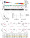
Fig. 1
Fig. 1. A large-scale trial using the PDX model and comprehensive molecular profiling.
a E7820 sensitivity, HRR gene alterations, and tumor mutation burden (TMB) of 42 PDXs. b Volcano plot representation of Wilcoxon’s rank sum test comparing the E7820 sensitivity of PDXs with and without mutations in 73 cancer-related genes that were mutated in at least five PDXs. c Scatter plots showing the E7820 response and gene expression. d The drug efficacy in the short term (3 days) or long term (12 days) was investigated. The viability of DLD1-P, DLD1-KO, HCC1937, and HCC1428 cells was evaluated after 3 days or 12 days of treatment with E7820 or olaparib. The IC50 values of E7820 and olaparib in nine cell lines on day 3 or day 12 are shown in tables.
Fig. 2
Fig. 2. BRCA2 dysfunction increases E7820-induced DNA double-strand breaks and sensitizes cells to E7820.
a γH2AX, a marker of DNA double-strand breaks, was evaluated by western blotting of cells treated with E7820 and olaparib. Treatment with 1 μM E7820 for 72 h degraded RBM39 protein and induced the appearance of more γH2AH than treatment with 1 μM olaparib. GAPDH was used as a loading control. b γH2AX induction in DLD1-P or DLD1-KO cells was evaluated by western blotting after 1 μM E7820 treatment. γH2AX induction first appeared 24 h after E7820 treatment and increased up to 120 h. GAPDH was used as a loading control. c DLD1 cells (P or KO) were treated with olaparib (10 µM), E7820 (1 µM), or indisulam (1 µM) for 72 h, and γH2AX, pRPA, and Rad51 foci were analyzed. Representative images are provided. NT no treatment; Scale bars in images, 10 µm. d The numbers of foci for γH2AX, pRPA, and Rad51 were counted and statistically analyzed using Two-tailed Welch’s t-test. Bars in the graph show means ± standard deviations. NT no treatment, E E7820, I indisulam, O olaparib. e Drug sensitivities to indisulam, E7820, and olaparib were investigated in DLD1-P and artificially generated DLD1 cell lines, in which genes were genetically engineered with a knockout (KO) of ATM, ATR, and BAP1 by the CRISPR‒Cas9 system. Sensitivity was assessed on day 12 after drug treatment.
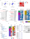
Fig. 3
Fig. 3. Transcriptomic changes associated with E7820.
a Transcriptomic changes associated with E7820. RNA-seq was performed in six cell lines to compare the changes in gene expression induced by E7820. The expression changes (log fold change; log FC) were highly concordant between DLD1-P and DLD1-KO cells (r = 0.95, left panel) or PC9 cells (r = 0.86, middle panel). The heatmap on the right shows that the trends in expression profile changes associated with E7820 were similar between any pair of DLD1-P, DLD1-KO, PC9, FUJI, H2228, and KMLS1. b The genes that were commonly upregulated (fold change > 1.1) and downregulated (fold change < 0.9) by E7820 treatment among the six cell lines included 1655 and 2787 genes, respectively. Ingenuity pathway analysis (IPA) was conducted for pathway analysis. Among the identified pathways shared by upregulated genes, “Protein Ubiquitination Pathway” and “Spliceosomal Cycles” were highly ranked (−log(p value) = 9.01 and 3.33, respectively). In contrast, the enriched pathways of the downregulated genes included pathways related to DNA damage repair, such as “Nucleotide excision repair (NER) Enhanced Pathway”, “Hereditary Breast Cancer Signaling”, and “Role of BRCA1 in DNA Damage Response” (−log(p value) = 9.27, 8.51 and 7.38, respectively). c The changes in gene expression induced by E7820 treatment are shown. The fold changes are shown in a heatmap comparing the expression of cell lines after E7820 treatment (1 μM for 48 h) to basal expression. The genes are involved in homologous recombination repair (HRR), nonhomologous end joining (NHEJ), the Fanconi anemia complementation group (FANC) pathway, nucleotide excision repair (NER), base excision repair (BER) and other pathways related to DNA damage repair (Common). Only genes whose expression was decreased by 10%< are shown. Information on all genes is shown in Supplemental Fig. 4. d The number of splicing anomalies induced by E7820 treatment is indicated. Similar alternative splicing events were induced by E7820 in both DLD1-P and DLD1-KO cells. Mis-splicing event codes are as follows: TSS transcription start site, A5SS alternative 5’ splice site, A3SS alternative 3’ splice site, SES single-exon skipping, MES multiple-exon skipping, IR intron retention, OR overlapping region. e The changes in IR ratio induced by E7820 treatment are shown. The most significant changes in individual genes of Fig. 3c are shown in a heatmap comparing the IR ratios of cell lines after E7820 treatment (1 μM for 48 h) to basal ratio. f Full-length cDNAs were selectively prepared from the cells and sequenced with a Sequel II long-read sequencer (Pacific Biosystems). The long-read sequences were used to identify isoforms using the bulk Iso-seq workflow and pigeon workflow. The isoforms were visualized using IGV (Integrative Genomics Viewer). The individual long reads were tagged with PB ID, as shown on the left. Intron retention within FANCD2 was specifically observed in the cells treated with E7820 (arrowheads). g The protein levels of FANCD2 and FANCA in DLD1-KO cells. DLD1-KO cells were incubated with E7820 for the indicated times, and western blotting was conducted to evaluate the protein expression of FANCD2, FANCA, γH2AX, and cleaved caspase-3.
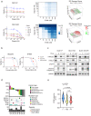
Fig. 4
Fig. 4. Combination treatment with E7820 and olaparib or cisplatin.
a Combination therapy with E7820 and olaparib. DLD1-P and DLD1-KO cells were treated with a combination of E7820 and olaparib at the indicated concentrations. Cell viability was assessed with a PrestoBlue cell viability assay. Synergistic effects are indicated in the 3D drug synergy maps. The surfaces of the maps were color-coded according to the ZIP synergy scores of the combination treatment. b The olaparib-resistant DLD1-KO cell line (DLD1-KO-OR) was developed through treatment with gradually increasing concentrations of olaparib (upto 20 μM). The resistant cell was treated with E7820 or olaparib for 12 days, and cell viability was assessed. c DLD1-P, DLD1-KO, and DLD1-KO-OR cells were treated with E7820 (1 μM), olaparib (1 μM), and their combination. The protein expression of the indicated genes was evaluated by western blotting. d PDXs with any HRR gene mutations were treated with E7820 (100 mg/kg). E7820 response (ΔT/C), HRR gene alterations, and TMB of 10 PDXs are shown. e A total of 52 PDX models were divided into three groups, and E7820 response (ΔT/C) were compared. (−) no mutations, VUS variant of unknown significance, PV pathogenic variants in HRR genes.

References

    1. Owa T, et al. Discovery of novel antitumor sulfonamides targeting G1 phase of the cell cycle. J. Med. Chem. 1999;42:3789–3799. doi: 10.1021/jm9902638. - DOI - PubMed
    1. Funahashi Y, et al. Sulfonamide derivative, E7820, is a unique angiogenesis inhibitor suppressing an expression of integrin alpha2 subunit on endothelium. Cancer Res. 2002;62:6116–6123. - PubMed
    1. Xu C, et al. RNA-binding protein 39: a promising therapeutic target for cancer. Cell Death Discov. 2021;7:214. doi: 10.1038/s41420-021-00598-7. - DOI - PMC - PubMed
    1. Han T, et al. Anticancer sulfonamides target splicing by inducing RBM39 degradation via recruitment to DCAF15. Science. 2017;356:eaal3755. doi: 10.1126/science.aal3755. - DOI - PubMed
    1. Uehara T, et al. Selective degradation of splicing factor CAPERalpha by anticancer sulfonamides. Nat. Chem. Biol. 2017;13:675–680. doi: 10.1038/nchembio.2363. - DOI - PubMed