

Surgical treatment of peri-implantitis
- PMID: 38789758
- PMCID: PMC11126382
- DOI: 10.1038/s41415-024-7405-9
Surgical treatment of peri-implantitis
Abstract
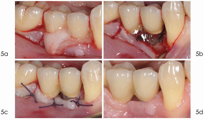
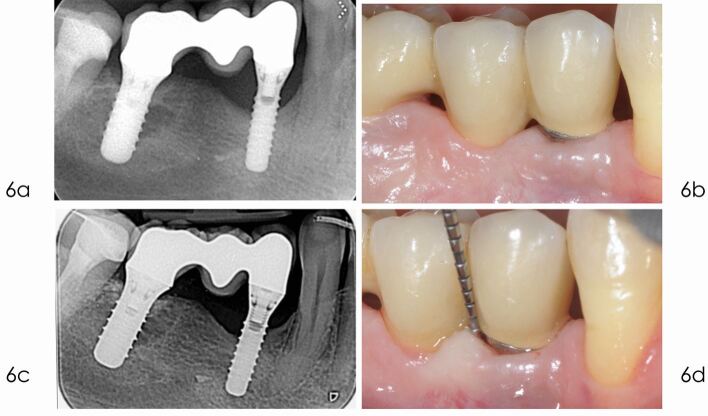
As utilisation of dental implants continues to rise, so does the incidence of biological complications. When peri-implantitis has already caused extensive bone resorption, the dentist faces the dilemma of which therapy is the most appropriate to maintain the implant. Since non-surgical approaches of peri-implantitis have shown limited effectiveness, the present paper describes different surgical treatment modalities, underlining their indications and limitations. The primary goal in the management of peri-implantitis is to decontaminate the surface of the infected implant and to eliminate deep peri-implant pockets. For this purpose, access flap debridement, with or without resective procedures, has shown to be effective in a large number of cases. These surgical treatments, however, may be linked to post-operative recession of the mucosal margin. In addition to disease resolution, reconstructive approaches also seek to regenerate the bone defect and to achieve re-osseointegration.
© 2024. The Author(s).
Conflict of interest statement
The authors declare no conflicts of interest.
Figures
Similar articles
-
Surgical interventions for the treatment of peri-implantitis.Clin Implant Dent Relat Res. 2023 Aug;25(4):682-695. doi: 10.1111/cid.13162. Epub 2022 Nov 23. Clin Implant Dent Relat Res. 2023. PMID: 36419243 Review.
-
Surgical therapy of peri-implantitis.Periodontol 2000. 2022 Feb;88(1):145-181. doi: 10.1111/prd.12417. Periodontol 2000. 2022. PMID: 35103328 Review.
-
Peri-implantitis: Summary and consensus statements of group 3. The 6th EAO Consensus Conference 2021.Clin Oral Implants Res. 2021 Oct;32 Suppl 21:245-253. doi: 10.1111/clr.13827. Clin Oral Implants Res. 2021. PMID: 34642987 Review.
-
Successful Management of Peri-Implantitis Using a Titanium Brush and a Doxycycline-Saline Slurry for Surface Detoxification With Guided Bone Regeneration: A 5-Year Follow-Up.Clin Adv Periodontics. 2020 Sep;10(3):118-122. doi: 10.1002/cap.10085. Epub 2020 Apr 10. Clin Adv Periodontics. 2020. PMID: 31845465
-
Re-osseointegration following reconstructive surgical therapy of experimental peri-implantitis. A pre-clinical in vivo study.Clin Oral Implants Res. 2019 May;30(5):447-456. doi: 10.1111/clr.13430. Epub 2019 May 3. Clin Oral Implants Res. 2019. PMID: 30963619
Cited by
-
Exploring Peri-Implantitis Risk-Factors: A Cross-Sectional Study.Dent J (Basel). 2025 Mar 28;13(4):148. doi: 10.3390/dj13040148. Dent J (Basel). 2025. PMID: 40277478 Free PMC article.
-
Erbium-Doped Yttrium Aluminium Garnet (Er:YAG) Lasers in the Treatment of Peri-Implantitis.Cureus. 2025 Jan 31;17(1):e78279. doi: 10.7759/cureus.78279. eCollection 2025 Jan. Cureus. 2025. PMID: 39897297 Free PMC article. Review.
-
Zirconium Dental Implants as Potential Optical Waveguides in Photodynamic Inactivation of Bacterial Biofilms-A Pilot Study.Microorganisms. 2025 Apr 8;13(4):850. doi: 10.3390/microorganisms13040850. Microorganisms. 2025. PMID: 40284686 Free PMC article.
-
Dysregulation of the Immune System in Advanced Periimplantitis: Systemic Implications and Inflammatory Mechanisms-A Hematological and Immunological Study.J Clin Med. 2025 Apr 3;14(7):2453. doi: 10.3390/jcm14072453. J Clin Med. 2025. PMID: 40217900 Free PMC article.
-
Progress in visible-light-activated photocatalytic coatings to combat implant-related infections: From mechanistic to translational roadmap.Bioact Mater. 2025 May 11;51:83-137. doi: 10.1016/j.bioactmat.2025.04.037. eCollection 2025 Sep. Bioact Mater. 2025. PMID: 40475082 Free PMC article. Review.
References
-
- Salvi G E, Stähli A, Imber J C et al. Physiopathology of peri-implant diseases. Clin Implant Dent Relat Res 2023; 25: 629-639. - PubMed
-
- Figuero E, Graziani F, Sanz I et al. Management of peri-implant mucositis and peri-implantitis. Periodontol 2000 2014; 66: 255-273. - PubMed
-
- Cosgarea R, Roccuzzo A, Jepsen K et al. Efficacy of mechanical/physical approaches for implant surface decontamination in non-surgical submarginal instrumentation of peri-implantitis. A systematic review. J Clin Periodontol 2023; 50: 188-211. - PubMed
-
- Herrera D, Berglundh T, Schwarz F et al. Prevention and treatment of peri-implant diseases - The EFP S3 level clinical practice guideline. J Clin Periodontol 2023; 50: 4-76. - PubMed
Publication types
MeSH terms
Substances
LinkOut - more resources
Full Text Sources
Research Materials
