Review
. 2024 May 2;11(5):451.
doi: 10.3390/bioengineering11050451.

New Frontiers in Breast Cancer Imaging: The Rise of AI

Affiliations
Review

New Frontiers in Breast Cancer Imaging: The Rise of AI

Stephanie B Shamir et al. Bioengineering (Basel). .

Abstract

Artificial intelligence (AI) has been implemented in multiple fields of medicine to assist in the diagnosis and treatment of patients. AI implementation in radiology, more specifically for breast imaging, has advanced considerably. Breast cancer is one of the most important causes of cancer mortality among women, and there has been increased attention towards creating more efficacious methods for breast cancer detection utilizing AI to improve radiologist accuracy and efficiency to meet the increasing demand of our patients. AI can be applied to imaging studies to improve image quality, increase interpretation accuracy, and improve time efficiency and cost efficiency. AI applied to mammography, ultrasound, and MRI allows for improved cancer detection and diagnosis while decreasing intra- and interobserver variability. The synergistic effect between a radiologist and AI has the potential to improve patient care in underserved populations with the intention of providing quality and equitable care for all. Additionally, AI has allowed for improved risk stratification. Further, AI application can have treatment implications as well by identifying upstage risk of ductal carcinoma in situ (DCIS) to invasive carcinoma and by better predicting individualized patient response to neoadjuvant chemotherapy. AI has potential for advancement in pre-operative 3-dimensional models of the breast as well as improved viability of reconstructive grafts.

Keywords: CNN; MRI; artificial intelligence; breast cancer; deep learning; mammography; risk stratification; ultrasound.


Conflict of interest statement

LRM—Medical Advisory Board ScreenPoint Medical.

Figures

Figure 1
Figure 1
Developing asymmetry detected by artificial intelligence (AI): Between the baseline screening mammogram (A) and the follow-up screening mammogram 17 months later (B), there has been a very subtle development of left breast asymmetry that is difficult to perceive with the naked eye. However, the AI program Transpara highlighted potential regions of interest (C) for the radiologist to query for additional mammographic and sonographic imaging. On further diagnostic imaging, the subtle asymmetry corresponds to a hypoechoic mass at left 4:00, 3 cm FN (D) with hypervascularity (E). AI program Koios correctly recognized the mass as “Probably Malignant”, and this area returned as a biopsy-proven invasive malignancy with lymphangitic spread (F). Images obtained from the Icahn School of Medicine at Mount Sinai.
Figure 1
Figure 1
Developing asymmetry detected by artificial intelligence (AI): Between the baseline screening mammogram (A) and the follow-up screening mammogram 17 months later (B), there has been a very subtle development of left breast asymmetry that is difficult to perceive with the naked eye. However, the AI program Transpara highlighted potential regions of interest (C) for the radiologist to query for additional mammographic and sonographic imaging. On further diagnostic imaging, the subtle asymmetry corresponds to a hypoechoic mass at left 4:00, 3 cm FN (D) with hypervascularity (E). AI program Koios correctly recognized the mass as “Probably Malignant”, and this area returned as a biopsy-proven invasive malignancy with lymphangitic spread (F). Images obtained from the Icahn School of Medicine at Mount Sinai.
Figure 2
Figure 2
New architectural distortion detected by artificial intelligence (AI): A patient in her 50s’ screening mammogram revealed a new area of architectural distortion (circle) in the inner central region of the left breast (A). The AI program Transpara highlighted potential regions of interest, including this suspicious area of architectural distortion on the left breast on the corresponding left CC view; however, AI also highlighted benign areas that were arbitrated out by the radiologist (B). There was no sonographic correlate, so a stereotactic biopsy of this area of architectural distortion was then biopsied under guidance. Pathology yielded invasive lobular carcinoma. Images obtained from the Icahn School of Medicine at Mount Sinai.
Figure 3
Figure 3
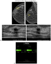
A new cancer diagnosis appropriately classified as “malignant” by artificial intelligence (AI): This patient in her 40s with a history of left breast carcinoma diagnosed 1 year prior, status post-left mastectomy with chemotherapy and hormonal therapy, presented with a palpable abnormality in the superficial lower outer left breast. No new or suspicious findings were seen on the patient’s diagnostic mammogram. Correlating with the patient’s concern about a palpable lump, diagnostic ultrasound revealed an irregularly shaped, hypoechoic mass with angular margins that are non-parallel (A), and Doppler shows no vascularity (B). The AI program Koios recognized this mass as “Probably Malignant” (C). This was returned as biopsy-proven invasive ductal carcinoma. Images obtained from the Icahn School of Medicine at Mount Sinai.
Figure 3
Figure 3
A new cancer diagnosis appropriately classified as “malignant” by artificial intelligence (AI): This patient in her 40s with a history of left breast carcinoma diagnosed 1 year prior, status post-left mastectomy with chemotherapy and hormonal therapy, presented with a palpable abnormality in the superficial lower outer left breast. No new or suspicious findings were seen on the patient’s diagnostic mammogram. Correlating with the patient’s concern about a palpable lump, diagnostic ultrasound revealed an irregularly shaped, hypoechoic mass with angular margins that are non-parallel (A), and Doppler shows no vascularity (B). The AI program Koios recognized this mass as “Probably Malignant” (C). This was returned as biopsy-proven invasive ductal carcinoma. Images obtained from the Icahn School of Medicine at Mount Sinai.
Figure 4
Figure 4
A benign finding appropriately classified as benign by artificial intelligence (AI): The patient initially presented for a bilateral screening mammogram and a bilateral screening breast ultrasound. A mammogram revealed benign dystrophic calcifications in the upper outer quadrant of the right breast (A). Correlating with findings on the mammogram, ultrasound revealed a complicated cyst showing posterior acoustic shadowing consistent with fat necrosis (B). The AI program Koios recognized this mass as “Benign” (C). Images obtained from the Icahn School of Medicine at Mount Sinai.
Figure 5
Figure 5
Artificial intelligence (AI) sequence utilized to accelerate image acquisition time: A deep resolve boost (DRB) AI sequence was utilized to increase the signal-to-noise ratio by artificially filling k-space, allowing for accelerated image acquisition. Images obtained from the Icahn School of Medicine at Mount Sinai.

References

    1. Hu Q., Giger M.L. Clinical Artificial Intelligence Applications: Breast Imaging. Radiol. Clin. N. Am. 2021;59:1027–1043. doi: 10.1016/j.rcl.2021.07.010. - DOI - PMC - PubMed
    1. Society A.C. Breast Cancer Facts & Figures 2022–2024. American Cancer Society, Inc.; Atlanta, GA, USA: 2022.
    1. Mango V.L., Sun M., Wynn R.T., Ha R. Should We Ignore, Follow, or Biopsy? Impact of Artificial Intelligence Decision Support on Breast Ultrasound Lesion Assessment. AJR Am. J. Roentgenol. 2020;214:1445–1452. doi: 10.2214/ajr.19.21872. - DOI - PMC - PubMed
    1. Ahn J.S., Shin S., Yang S.A., Park E.K., Kim K.H., Cho S.I., Ock C.Y., Kim S. Artificial Intelligence in Breast Cancer Diagnosis and Personalized Medicine. J. Breast Cancer. 2023;26:405–435. doi: 10.4048/jbc.2023.26.e45. - DOI - PMC - PubMed
    1. Coffey K., Aukland B., Amir T., Sevilimedu V., Saphier N.B., Mango V.L. Artificial Intelligence Decision Support for Triple-Negative Breast Cancers on Ultrasound. J. Breast Imaging. 2024;6:33–44. doi: 10.1093/jbi/wbad080. - DOI - PMC - PubMed

LinkOut - more resources