Review
. 2024 May 11;11(5):582.
doi: 10.3390/children11050582.

Congenital Long QT Syndrome in Children and Adolescents: A General Overview

Affiliations
Review

Congenital Long QT Syndrome in Children and Adolescents: A General Overview

Elia Balestra et al. Children (Basel). .

Abstract

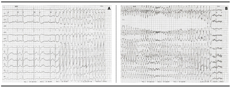
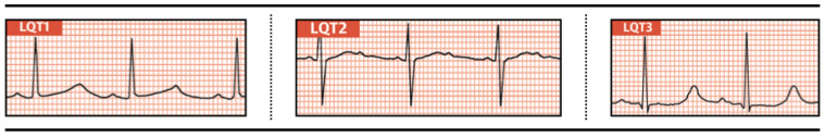
Congenital long QT syndrome (LQTS) represents a disorder of myocardial repolarization characterized by a prolongation of QTc interval on ECG, which can degenerate into fast polymorphic ventricular arrhythmias. The typical symptoms of LQTS are syncope and palpitations, mainly triggered by adrenergic stimuli, but it can also manifest with cardiac arrest. At least 17 genotypes have been associated with LQTS, with a specific genotype-phenotype relationship described for the three most common subtypes (LQTS1, -2, and -3). β-Blockers are the first-line therapy for LQTS, even if the choice of the appropriate patients needing to be treated may be challenging. In specific cases, interventional measures, such as an implantable cardioverter-defibrillator (ICD) or left cardiac sympathetic denervation (LCSD), are useful. The aim of this review is to highlight the current state-of-the-art knowledge on LQTS, providing an updated picture of possible diagnostic algorithms and therapeutic management.

Keywords: QTc interval; arrhythmias; congenital long QT syndrome; implantable cardioverter-defibrillator; left cardiac sympathetic denervation; palpitations; syncope; β-blockers.


Conflict of interest statement

The authors declare no conflicts of interest.

Figures

Figure 1
Figure 1
Action potential and electrical currents—Upper figure: inward and outward currents and their relationship with action potential phases; lower figure: electrocardiogram aligned in time with the corresponding action potential. INa—Sodium inward current; ICa-L—Slow calcium inward current; IKr, IKs, IK1—Rectifier potassium current; 0—Phase 0 or depolarization phase of action potential; 1—Phase 1 or early repolarization phase of action potential; 2—Phase 2 or plateau phase of action potential; 3—Phase 3 or late repolarization phase of action potential; 4—Phase 4 or resting phase of action potential.
Figure 2
Figure 2
Torsades des pointes and self-limiting ventricular fibrillation. In some cases malignant arrhythmias can be self-limiting; in this case, frequent ventricular extrasystoles are seen, the last of which triggers torsades des pointes (A), which degenerates into ventricular fibrillation (B) that ends spontaneously.
Figure 3
Figure 3
Genotype to electrical phenotype—LQT1: normal T waveform with large base implant; the wave’s amplitude could be low, normal, or high. LQT2: usually low-amplitude T waves with a notched shape (should not to be confused with U waves). LQT3: the main feature is the late onset of the T wave from the isoelectric line; therefore, most of the prolongation is given by the stretch between the Q wave and the beginning of the T wave, rather than by the T wave itself.
Figure 4
Figure 4
The maximum slope intercept method—The end of the T wave is defined by the intercept between the tangent drawn through the maximum downward slope of the T wave and the isoelectric line. (A) U wave is not included in the QT calculation; (B) U wave is fused to the T wave, so it’s included in the QT measurement and the tangent is drawn through the second slope; (C) The tangent is drawn through the maximum downward slope of the T wave and small U wave is not included in the QT calculation. Green line—isoelectric line; red line—tangent line to the maximum downward slope of the T wave; blue lines—indicating the beginning of QRS and the intercept between the tangent and isoelectric line; QT correspond to the interval between the two blue lines.
Figure 5
Figure 5
Algorithm for management of long QT syndrome patients (adapted from 2022 ESC Guidelines): ICD—implantable cardioverter-defibrillator; LCSD—left cardiac sympathetic denervation.

References

    1. Schwartz P.J., Ackerman M.J., Antzelevitch C., Bezzina C.R., Borggrefe M., Cuneo B.F., Wilde A.A.M. Inherited cardiac arrhythmias. Nat. Rev. Dis. Primers. 2020;6:58. doi: 10.1038/s41572-020-0188-7. - DOI - PMC - PubMed
    1. Schwartz P.J., Crotti L., Insolia R. Long-QT syndrome from genetics to management. Circ. Arrhythmia Electrophysiol. 2012;5:868–877. doi: 10.1161/CIRCEP.111.962019. - DOI - PMC - PubMed
    1. Schwartz P.J., Stramba-Badiale M., Crotti L., Pedrazzini M., Besana A., Bosi G., Gabbarini F., Goulene K., Insolia R., Mannarino S., et al. Prevalence of the congenital long-qt syndrome. Circulation. 2009;120:1761–1767. doi: 10.1161/CIRCULATIONAHA.109.863209. - DOI - PMC - PubMed
    1. Wilde A.A.M., Amin A.S., Postema P.G. Diagnosis, management and therapeutic strategies for congenital long QT syndrome. Heart. 2022;108:332–338. doi: 10.1136/heartjnl-2020-318259. - DOI - PMC - PubMed
    1. Asatryan B., Murray B., Gasperetti A., McClellan R., Barth A.S. Unraveling Complexities in Genetically Elusive Long QT Syndrome. Circ. Arrhythm. Electrophysiol. 2024;17:e012356. doi: 10.1161/CIRCEP.123.012356. - DOI - PubMed

LinkOut - more resources