. 2024 May 10;13(5):589.
doi: 10.3390/antiox13050589.

The Activity of YCA1 Metacaspase Is Regulated by Reactive Sulfane Sulfur via Persulfidation in Saccharomyces cerevisiae

Affiliations

The Activity of YCA1 Metacaspase Is Regulated by Reactive Sulfane Sulfur via Persulfidation in Saccharomyces cerevisiae

Qingda Wang et al. Antioxidants (Basel). .

Abstract

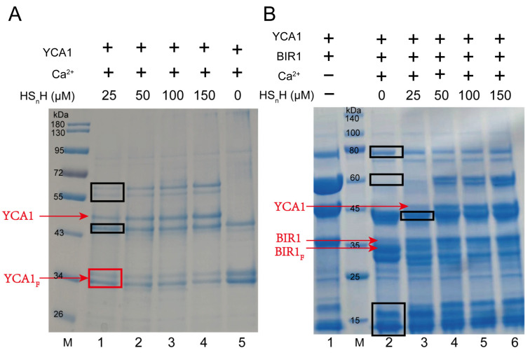
YCA1, the only metacaspase in Saccharomyces cerevisiae, plays important roles in the regulation of chronological lifespan, apoptosis, and cytokinesis. YCA1 has protein hydrolase activity and functions by cleaving itself and target proteins. However, there are few reports about the regulation of YCA1 activity. In this study, we observed that reactive sulfane sulfur (RSS) can inhibit the activity of YCA1. In vitro experiments demonstrated that RSS reacted with the Cys276 of YCA1, the residue central to its protein hydrolase activity, to form a persulfidation modification (protein-SSH). This modification inhibited both its self-cleavage and the cleavage of its substrate protein, BIR1. To investigate further, we constructed a low-endogenous-RSS mutant of S. cerevisiae, BY4742 Δcys3, in which the RSS-producing enzyme cystathionine-γ-lyase (CYS3) was knocked out. The activity of YCA1 was significantly increased by the deletion of CYS3. Moreover, increased YCA1 activity led to reduced chronological lifespan (CLS) and CLS-driven apoptosis. This study unveils the first endogenous factor that regulates YCA1 activity, introduces a novel mechanism of how yeast cells regulate chronological lifespan, and broadens our understanding of the multifaceted roles played by RSS.

Keywords: YCA1; apoptosis; chronological lifespan; metacaspase; persulfidation; reactive sulfane sulfur.


Conflict of interest statement

The authors declare no conflicts of interest.

Figures

Figure 1
Figure 1
RSS inhibited self-cleavage and proteolytic activity of YCA1. (A) YCA1 at 5 μM was added to the reaction buffer. CaCl2 was added at a concentration of 10 mM. Different concentrations of HSnH were added (Lanes 1–4, 25 μM–150 μM). Lane 5 (no HSnH addition) was the control. Uncleaved YCA1 and cleaved YCA1 are labeled with red lines. YCA1F represents cleaved YCA1. M represents the protein marker. (B) Purified YCA1 at 5 μM and BIR1 at 5 μM were mixed in the reaction buffer. Lane 1 contains only YCA1 and BIR1. Lane 2 contains YCA1, BIR1, and Ca2+ but no HSnH. CaCl2 (10 mM) was added in lanes 3~6. HSnH at 25 μM–150 μM were added in lanes 3~6. BIR1F represents cleaved BIR1. M represents the protein marker. Contaminating proteins co-purified with YCA1 from E. coli are labelled with black boxes.
Figure 2
Figure 2
YCA1 was modified by RSS at the Cys276 position. (A) The crystal structure of YCA1. The data were obtained from PDB ID: 4f6o and were processed with PyMOL (2.0.7). (B) The MS2 spectra of the Cys276-containing peptide from the HSnH-reacted YCA1. “C-S-AM” represents the thiol group of Cys276 (Cys-SH) modified by persulfidation to form Cys-SSH, which is then blocked by IAM to form Cys-SS-AM. (C) The MS2 spectra of the Cys276-containing peptide from the DTT-treated YCA1. “C-AM” represents the thiol group of Cys276 (Cys-SH) blocked by IAM to form Cys-S-AM. The peptide fragments generated by the y, b ions are labeled in blue and red, respectively.
Figure 3
Figure 3
Knocking out cys3 reduces intracellular RSS levels. (A) The BY4742 wt and Δcys3 strains were cultivated in YPD liquid medium. (B) The intracellular RSS levels of the BY4742 wt and Δcys3 at different times when cultivated in YPD medium. “*” represents a difference (p < 0.05), “**” represents a significant difference (p < 0.01), “***” represents a significant difference (p < 0.005) and “ns” represents no significant differences (p > 0.05) in the two-sided t-test. The data are from three replicated experiments and are shown as averages ± s.d.
Figure 4
Figure 4
The activity of YCA1 of cleaving BIR1 was inhibited in Δcys3. (A) The Western blot analysis of the intracellular fragments of BIR1-GFP. The BIR1-GFP cleaved fragments are indicated with red lines. “CK” represents BY4742 cells without BIR1-GFP. (B) Changes in the transcriptional levels of YCA1 and BIR1 in Δcys3, with the wt as the control. The amounts of gene transcripts were from the transcriptomic analysis. The fold change was calculated by dividing the transcript amount in Δcys3 by that in the wt. “ns” represents no differences (p > 0.05) in the two-sided t-test. The data in (B) are from three replicated experiments and are shown as averages ± s.d.
Figure 5
Figure 5
Increasing YCA1 activity resulted in physiological changes in S. cerevisiae. (A) The CLS of Δcys was shorter than that of the wt and Δcys3Δyca1. (BD) The apoptosis status of BY4742 wt, Δcys3, and Δcys3Δyca1 cells was analyzed by V-FITC/PI double staining after cultivation in SD medium for 72 h. The data in (A) are from three replicated experiments and are shown as averages ± s.d. The cell numbers are labeled in different colors, with red representing the highest cell count.
Figure 6
Figure 6
The transcriptomics analysis of the BY4742 wt and Δcys3 strains. (A) The number of genes changed significantly at the transcriptional level. (B) Heat shock protein genes were significantly up-regulated in Δcys3. (C) Carbon metabolism-related genes were significantly up-regulated in Δcys3. For each strain, three samples were collected and analyzed in parallel.

References

    1. Liu Z.P., Ji Q.Z., Ren J., Yan P.Z., Wu Z.M., Wang S., Sun L., Wang Z.H., Li J.M., Sun G.Q., et al. Large-scale chromatin reorganization reactivates placenta-specific genes that drive cellular aging. Dev. Cell. 2022;57:1347–1368.e12. doi: 10.1016/j.devcel.2022.05.004. - DOI - PubMed
    1. Zhang L., Pitcher L.E., Yousefzadeh M.J., Niedernhofer L.J., Robbins P.D., Zhu Y. Cellular senescence: A key therapeutic target in aging and diseases. J. Clin. Investig. 2022;132:e158450. doi: 10.1172/JCI158450. - DOI - PMC - PubMed
    1. Murphy B.M., Burd C.E. The aging lung microenvironment awakens melanoma metastases. Cancer Cell. 2022;40:815–817. doi: 10.1016/j.ccell.2022.06.013. - DOI - PubMed
    1. Priault M., Camougrand N., Kinnally K.W., Vallette F.M., Manon S. Yeast as a tool to study Bax/mitochondrial interactions in cell death. FEMS Yeast Res. 2003;4:15–27. doi: 10.1016/S1567-1356(03)00143-0. - DOI - PubMed
    1. Kaya A., Lobanov A.V., Gladyshev V.N. Evidence that mutation accumulation does not cause aging in Saccharomyces cerevisiae. Aging Cell. 2015;14:366–371. doi: 10.1111/acel.12290. - DOI - PMC - PubMed

LinkOut - more resources