. 2024 May 12;25(10):5272.
doi: 10.3390/ijms25105272.

miRNA Expression Profiles in Isolated Ventricular Cardiomyocytes: Insights into Doxorubicin-Induced Cardiotoxicity

Affiliations

miRNA Expression Profiles in Isolated Ventricular Cardiomyocytes: Insights into Doxorubicin-Induced Cardiotoxicity

Yohana Domínguez Romero et al. Int J Mol Sci. .

Abstract

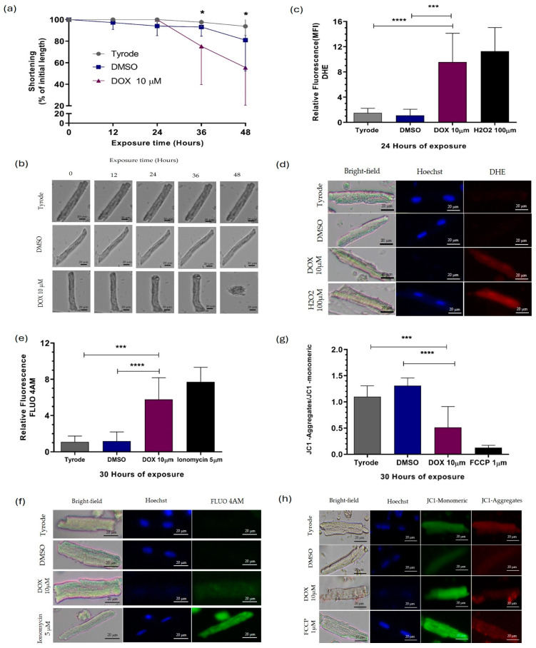
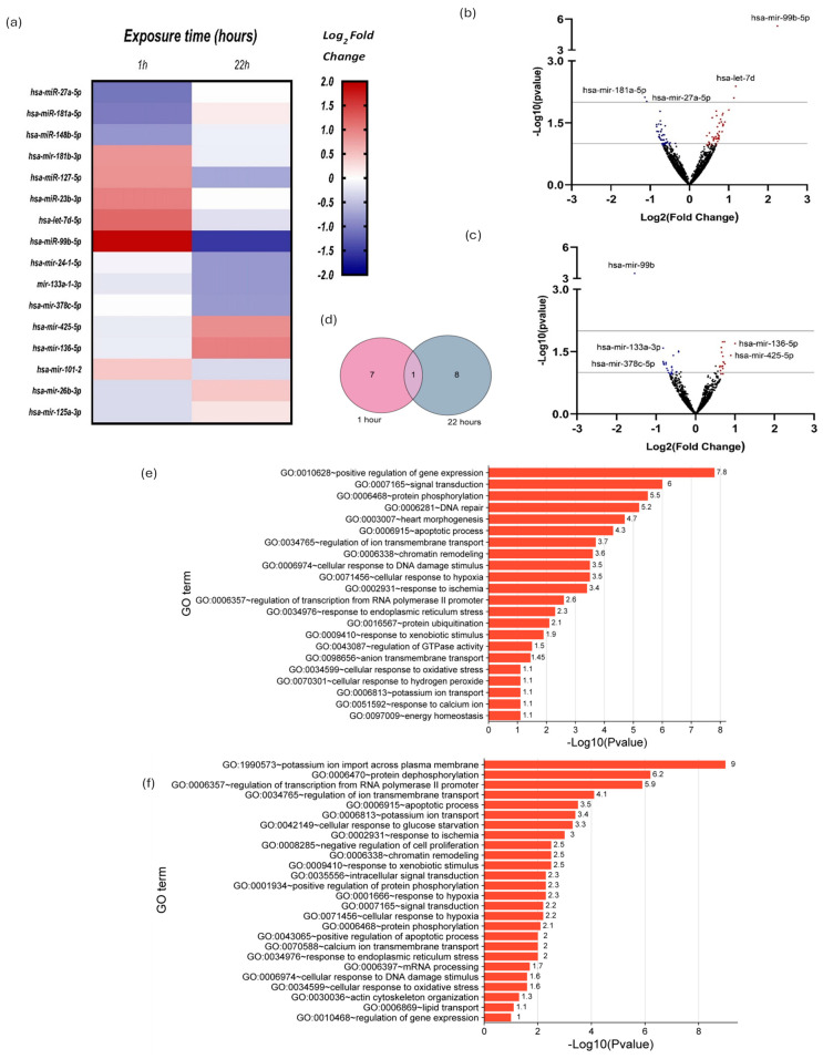
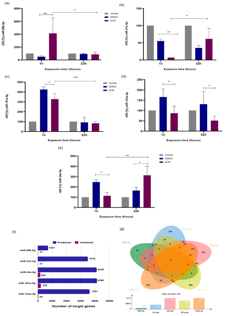
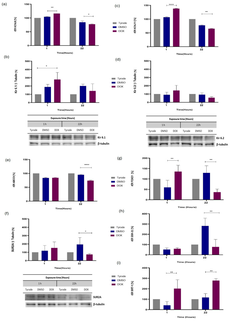
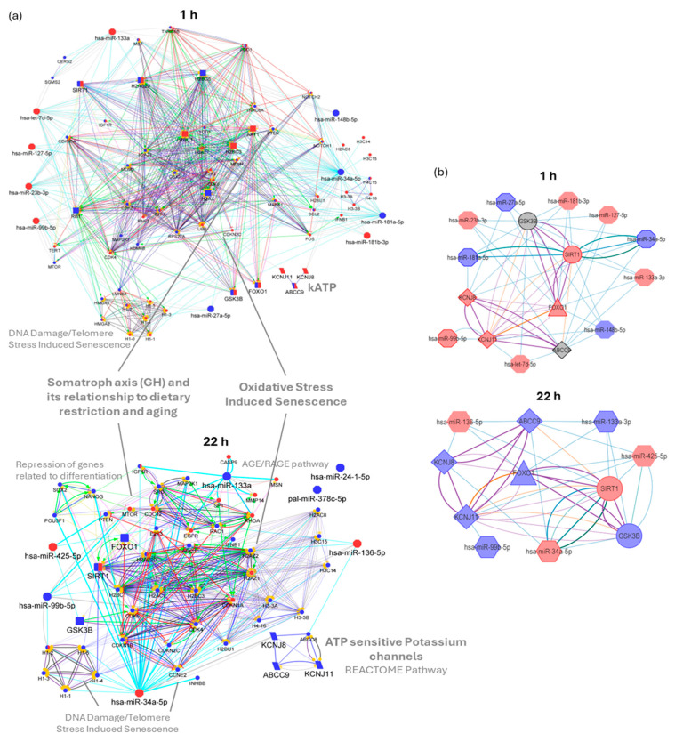
Doxorubicin (DOX), widely used as a chemotherapeutic agent for various cancers, is limited in its clinical utility by its cardiotoxic effects. Despite its widespread use, the precise mechanisms underlying DOX-induced cardiotoxicity at the cellular and molecular levels remain unclear, hindering the development of preventive and early detection strategies. To characterize the cytotoxic effects of DOX on isolated ventricular cardiomyocytes, focusing on the expression of specific microRNAs (miRNAs) and their molecular targets associated with endogenous cardioprotective mechanisms such as the ATP-sensitive potassium channel (KATP), Sirtuin 1 (SIRT1), FOXO1, and GSK3β. We isolated Guinea pig ventricular cardiomyocytes by retrograde perfusion and enzymatic dissociation. We assessed cell morphology, Reactive Oxygen Species (ROS) levels, intracellular calcium, and mitochondrial membrane potential using light microscopy and specific probes. We determined the miRNA expression profile using small RNAseq and validated it using stem-loop qRT-PCR. We quantified mRNA levels of some predicted and validated molecular targets using qRT-PCR and analyzed protein expression using Western blot. Exposure to 10 µM DOX resulted in cardiomyocyte shortening, increased ROS and intracellular calcium levels, mitochondrial membrane potential depolarization, and changes in specific miRNA expression. Additionally, we observed the differential expression of KATP subunits (ABCC9, KCNJ8, and KCNJ11), FOXO1, SIRT1, and GSK3β molecules associated with endogenous cardioprotective mechanisms. Supported by miRNA gene regulatory networks and functional enrichment analysis, these findings suggest that DOX-induced cardiotoxicity disrupts biological processes associated with cardioprotective mechanisms. Further research must clarify their specific molecular changes in DOX-induced cardiac dysfunction and investigate their diagnostic biomarkers and therapeutic potential.

Keywords: FOXO1; GSK3B; KATP; SIRT1; cardioprotection; cardiotoxicity; doxorubicin; miRNAs.


Conflict of interest statement

The authors declare no conflicts of interest.

Figures

Figure 1
Figure 1
DOX-induced injury in ventricular cardiomyocytes of Cavia porcellus. We exposed isolated cardiomyocytes to 10 μM DOX: (a) % shortening and (b) representative photographs of the shortening of cardiomyocytes during 48 h of exposure to DOX, (c) ROS levels analyzed with DHE, (d) representative photographs of ROS production in cardiomyocytes exposed to DOX for 22 h, (e) intracellular calcium levels analyzed with FLU4AM and (f) representative photographs of calcium levels in cardiomyocytes exposed to DOX for 30 h. (g) Mitochondrial membrane potential (ΔΨm) analyzed with JC1 and (h) representative photographs of ΔΨm in cardiomyocytes exposed to DOX for 30 h. Nuclear counterstain with DAPI (blue), quantification of expression by mean fluorescence intensity (MFI), Data were analyzed using two-way ANOVA parametric statistical test (p < 0.05, n = 10 cardiomyocytes per group)—representative graphs of three different experiments. Significance level of statistical findings: (*) p < 0.0332, (***) p < 0.0002, significant (****) p < 0.0001.
Figure 2
Figure 2
The expression miRNAs from Cavia porcellus isolated ventricular cardiomyocytes exposed to 10 µM DOX for 1 and 22 h. (a) Heat map of 16 expressed miRNAs obtained by small RNA-seq versus vehicle control cells (DMSO); red, expression up; blue, expression down; and white, indicator of no variation. Differential expression by using the DESeq2 program. (b,c) Volcano plots of miRNAs from cardiomyocytes exposed to 10 µM DOX for 1 and 22 h, respectively. Blue dots represent downregulated miRNAs, and red dots represent upregulated miRNAs. We only included miRNAs with –log10 (p value) ≥ 1 and log2 fold change ≥ 0.8 and ≤ −0.8 in blue or red dots. (d) Venn diagrams showing the numbers of miRNAs differentially expressed at 1 h and 22 h. (e,f) The related biological process with DOX-induced cardiotoxicity –log10 (p value) ≥ 1.
Figure 3
Figure 3
Validation of the differential expression of some miRNAs associated with Cavia porcellus ventricular cardiomyocytes’ response to DOX-induced injury. Relative expression ratios (rERs) of miRNAs by RT-qPCR stem-loop for (a) miR-99b-5p, (b) miR-27a-5p, (c) miR-181a-5p, (d) miR-133a-3p, and (e) miR-34a-5p (n = 3). (f) Several target genes predicted and validated to miRNAs are evaluated. (g) Venn diagrams showing the numbers of target genes predicted and validated shared between the miRNAs analyzed. Data were analyzed using a two-way ANOVA parametric statistical test (p < 0.05, n = 3)—representative graphs of three experiments. Significance level of statistical findings: (*) p < 0.0332, (**) p < 0.0021 (***) p < 0.0002. We performed target gene analysis in CSmiRTar. (http://cosbi4.ee.ncku.edu.tw/CSmiRTar/search) (accessed on 27 March 2024). At least two databases support the predicted target genes, and the average normalized score (ANS) is ≥0.2.
Figure 4
Figure 4
The expression of genes and their protein products is associated with Cavia porcellus ventricular cardiomyocytes’ response to DOX-induced injury. Relative expression radius (rER) of target genes (a) KCNJ8, (c) KCNJ11, (e) ABCC9, (g) FOXO1, (h) GSK3β, and (i) SIRT-1 by RT-qPCR and protein products (b) Kir 6.1, (d) Kir 6.2, and (f) SUR2A by Western blot. Data were analyzed using a two-way ANOVA parametric statistical test (p < 0.05, n = 3)—representative graphs of three experiments. Significance level of statistical findings: (*) p < 0.0332, (**) p < 0.0021, significant (****) p < 0.0001.
Figure 5
Figure 5
miRNA–target interactions (MTIs) and protein–protein interaction (PPI) network for DOX-altered miRNAs and genes. (a) CluePedia visualization of miRNA, proteins of interest, and relevant validated target genes obtained from miRecord and mirTarBase. (b) Summary network of experimentally evaluated miR and genes, where thick border node represents confirmation by RT-PCR. Blue and red nodes correspond to downregulated and upregulated molecules, respectively. The label font size and node size highlight the selected molecules. Target genes from phenotype-relevant pathways in circles, miRNAs in octagons, cardioprotective-related proteins in rounded rectangles (a) or circles (b), network-relevant proteins (degree ≥ 4) in rectangles, KATP channel subunits in parallelograms (a) or diamonds (b). Edge width is proportional to Kappa score and color edges depict interaction type: (a) predicted MTI in grey, validated MTI in aquamarine (miRTarBase) and blue (miRecords), activation in green, binding in blue, catalysis in purple, expression in yellow, inhibition in red, reaction in black, PTM in fuchsia. For (b), color edges depict interaction source: CSmiRTar in blue, validated miRTarbase and miRecord in aquamarine and green, respectively, PPI STRING in purple, and literature in orange. Networks were built with Cytoscape 3.9.1.

Similar articles

Cited by

References

    1. Müller I., Jenner A., Bruchelt G., Niethammer D., Halliwell B. Effect of Concentration on the Cytotoxic Mechanism of Doxorubicin-Apoptosis and Oxidative DNA Damage. Biochem. Biophys. Res. Commun. 1997;230:254–257. doi: 10.1006/bbrc.1996.5898. - DOI - PubMed
    1. Carvalho C., Santos R., Cardoso S., Correia S., Oliveira P., Santos M., Moreira P. Doxorubicin: The Good, the Bad and the Ugly Effect. Curr. Med. Chem. 2009;16:3267–3285. doi: 10.2174/092986709788803312. - DOI - PubMed
    1. Sheibani M., Azizi Y., Shayan M., Nezamoleslami S., Eslami F., Farjoo M.H., Dehpour A.R. Doxorubicin-Induced Cardiotoxicity: An Overview on Pre-Clinical Therapeutic Approaches. Cardiovasc. Toxicol. 2022;22:292–310. doi: 10.1007/S12012-022-09721-1. - DOI - PubMed
    1. Octavia Y., Tocchetti C.G., Gabrielson K.L., Janssens S., Crijns H.J., Moens A.L. Doxorubicin-Induced Cardiomyopathy: From Molecular Mechanisms to Therapeutic Strategies. J. Mol. Cell Cardiol. 2012;52:1213–1225. doi: 10.1016/j.yjmcc.2012.03.006. - DOI - PubMed
    1. Carvalho F.S., Burgeiro A., Garcia R., Moreno A.J., Carvalho R.A., Oliveira P.J. Doxorubicin-Induced Cardiotoxicity: From Bioenergetic Failure and Cell Death to Cardiomyopathy. Med. Res. Rev. 2014;34:106–135. doi: 10.1002/med.21280. - DOI - PubMed

MeSH terms