Review
. 2024 May 14;25(10):5349.
doi: 10.3390/ijms25105349.

Electro-Metabolic Coupling of Cumulus-Oocyte Complex

Affiliations
Review

Electro-Metabolic Coupling of Cumulus-Oocyte Complex

Diletta Del Bianco et al. Int J Mol Sci. .

Abstract

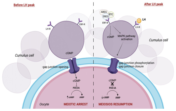
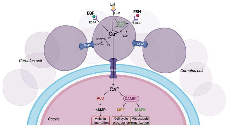
Oocyte-cumulus cell interaction is essential for oocyte maturation and competence. The bidirectional crosstalk network mediated by gap junctions is fundamental for the metabolic cooperation between these cells. As cumulus cells exhibit a more glycolytic phenotype, they can provide metabolic substrates that the oocyte can use to produce ATP via oxidative phosphorylation. The impairment of mitochondrial activity plays a crucial role in ovarian aging and, thus, in fertility, determining the success or failure of assisted reproductive techniques. This review aims to deepen the knowledge about the electro-metabolic coupling of the cumulus-oocyte complex and to hypothesize a putative role of potassium channel modulators in order to improve fertility, promote intracellular Ca2+ influx, and increase the mitochondrial biogenesis and resulting ATP levels in cumulus cells.

Keywords: cumulus cells; gap junctions; glucose metabolism; oocyte; resveratrol.


Conflict of interest statement

The authors declare no conflicts of interest.

Figures

Figure 1
Figure 1
Bidirectional cumulus–oocyte relationship. A model showing intercellular communication in the cumulus–oocyte complex (COC). The cellular crosstalk between the oocyte and the surrounding somatic cells is mediated by communication through gap junctions, allowing the passage of low-molecular-weight molecules. Created with BioRender.com (accessed on 25 January 2024).
Figure 2
Figure 2
Regulation of oocyte maturation. Schematic diagram of oocyte meiotic arrest in the phase preceding the gonadotropic peak (left) and gonadotropin-induced oocyte meiotic resumption (right). Meiotic regulation is modulated by the levels of cyclic guanosine monophosphate (cGMP) and Adenosine 3′,5′-cyclic monophosphate (cAMP) that are transferred from cumulus cells (CCs) to the oocyte. Created with BioRender.com (accessed on 25 January 2024).
Figure 3
Figure 3
Cumulus–oocyte metabolic coupling. Cumulus and oocyte cell metabolic reprogramming is dependent on the pyruvate dehydrogenase kinase (PDK) and pyruvate dehydrogenase (PDH) enzymes. The schematic illustration shows the molecular mechanisms of glucose metabolism within the COC. Metabolic cooperation between the two cell types is also made possible by the transfer of Adenosine triphosphate (ATP) generated by the heap via glycolysis and the adenosine salvage pathway. Created with BioRender.com (accessed on 25 January 2024).
Figure 4
Figure 4
Mitochondrial biogenesis. The schematic diagram shows how resveratrol decreases the functional expression of voltage-dependent potassium currents by causing a depolarization of the cell membrane in human ovarian granulosa cells (hGCs). This event promotes an increase in intracellular Ca2+ that leads to an improvement in mitochondrial function with an increase in mitochondrial biogenesis. Created with BioRender.com (accessed on 25 January 2024).
Figure 5
Figure 5
Role of intracellular calcium in oocyte maturation. Follicle-stimulating hormone (FSH), luteinizing hormone (LH), and epidermal growth factor-like (EGF-like) paracrine factors bind to their receptors on the cumulus cell and induce intracellular Ca mobilization. The increase of intracellular Ca2+ in the CCs can be transmitted to the oocyte through gap junctions. In the oocyte, Ca can inhibit adenylyl cyclase isoform 3 (AC3), resulting in a reduction in cAMP in the oocyte. Alternatively, Ca2+ can activate Ca/calmodulin-dependent protein kinase II (CAMKII), which in turn activates Maturation Promoting Factor (MPF) and mitogen-activated protein kinase (MAPK), promoting cell cycle progression and spindle formation important for oocyte maturation. Created with BioRender.com.
Figure 6
Figure 6
Metabolic switch in COC depending on the stage of folliculogenesis. The schematic diagram shows the metabolic switch of the COC in relation to follicle size depending on the stage of folliculogenesis. However, the metabolism of CCs is oxidative when the follicle is small and well irrigated, while it becomes glycolytic when the follicle grows and matures. Cx43 levels during the folliculogenesis indicate the formation of the maximum number of gap junctions when the follicle is mature. Created with BioRender.com (accessed on 25 January 2024).

Similar articles

Cited by

References

    1. Eppig J.J., Schultz R.M., O’Brien M., Chesnel F. Relationship between the Developmental Programs Controlling Nuclear and Cytoplasmic Maturation of Mouse Oocytes. Dev. Biol. 1994;164:1–9. doi: 10.1006/dbio.1994.1175. - DOI - PubMed
    1. Hussein T.S., Thompson J.G., Gilchrist R.B. Oocyte-Secreted Factors Enhance Oocyte Developmental Competence. Dev. Biol. 2006;296:514–521. doi: 10.1016/j.ydbio.2006.06.026. - DOI - PubMed
    1. Fontana J., Martínková S., Petr J., Žalmanová T., Trnka J. Metabolic Cooperation in the Ovarian Follicle. Physiol. Res. 2020;69:33–48. doi: 10.33549/physiolres.934233. - DOI - PMC - PubMed
    1. Gershon E., Plaks V., Dekel N. Gap Junctions in the Ovary: Expression, Localization and Function. Mol. Cell. Endocrinol. 2008;282:18–25. doi: 10.1016/j.mce.2007.11.001. - DOI - PubMed
    1. Liu Y.-X., Zhang Y., Li Y.-Y., Liu X.-M., Wang X.-X., Zhang C.-L., Hao C.-F., Deng S.-L. Regulation of Follicular Development and Differentiation by Intra-Ovarian Factors and Endocrine Hormones. Front. Biosci.-Landmark. 2019;24:983–993. doi: 10.2741/4763. - DOI - PubMed