. 2024 May 15;25(10):5403.
doi: 10.3390/ijms25105403.

BMPR2 Loss Activates AKT by Disrupting DLL4/NOTCH1 and PPARγ Signaling in Pulmonary Arterial Hypertension

Affiliations

BMPR2 Loss Activates AKT by Disrupting DLL4/NOTCH1 and PPARγ Signaling in Pulmonary Arterial Hypertension

Keytam S Awad et al. Int J Mol Sci. .

Abstract

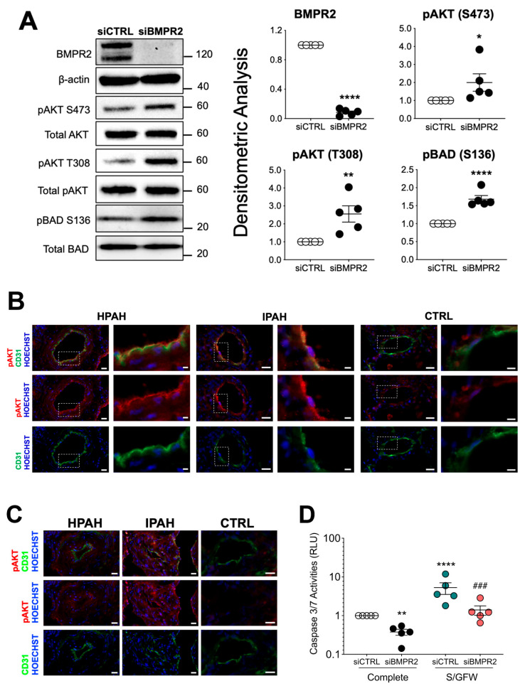
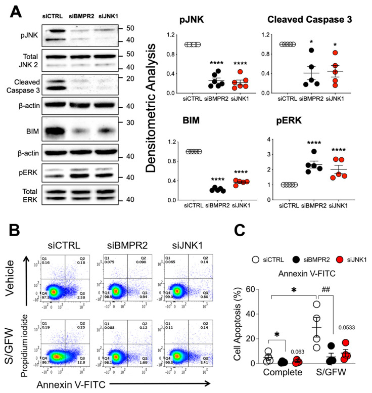
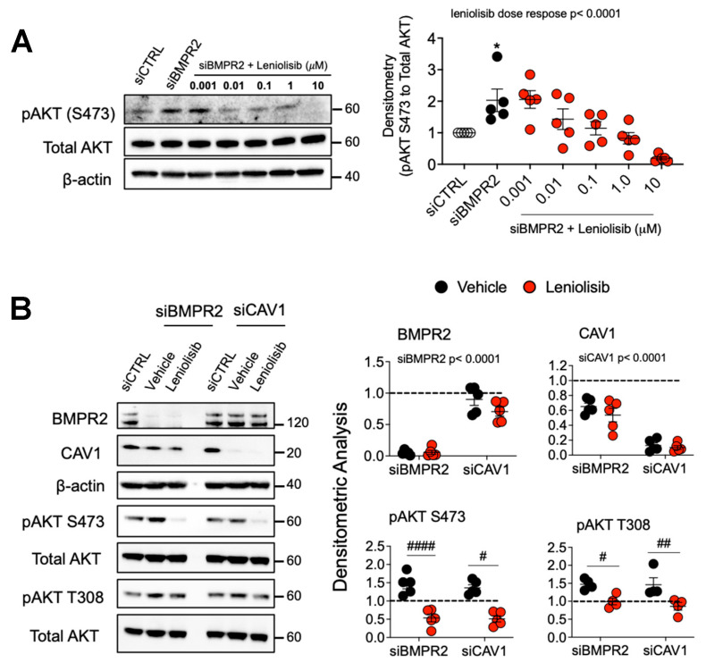
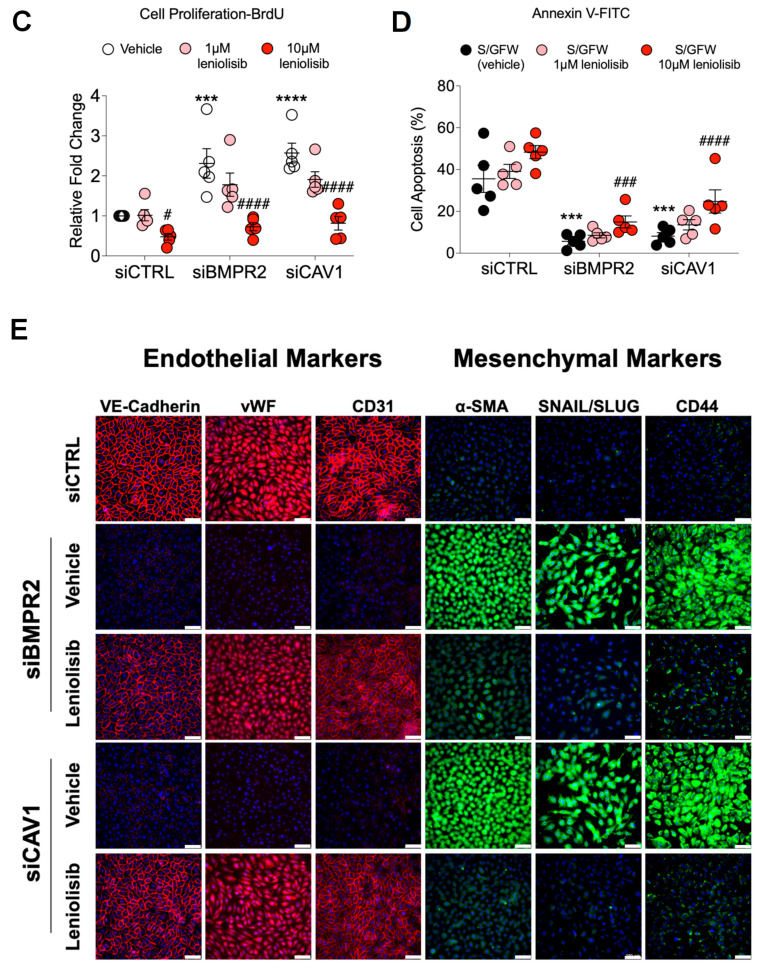
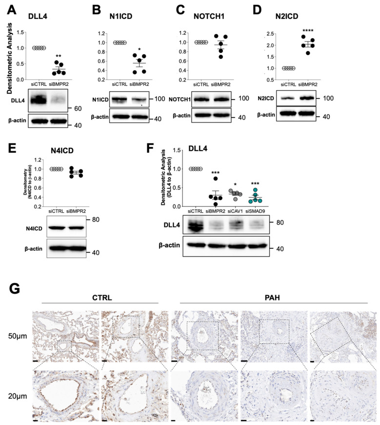
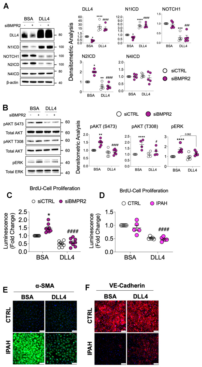
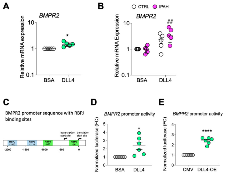
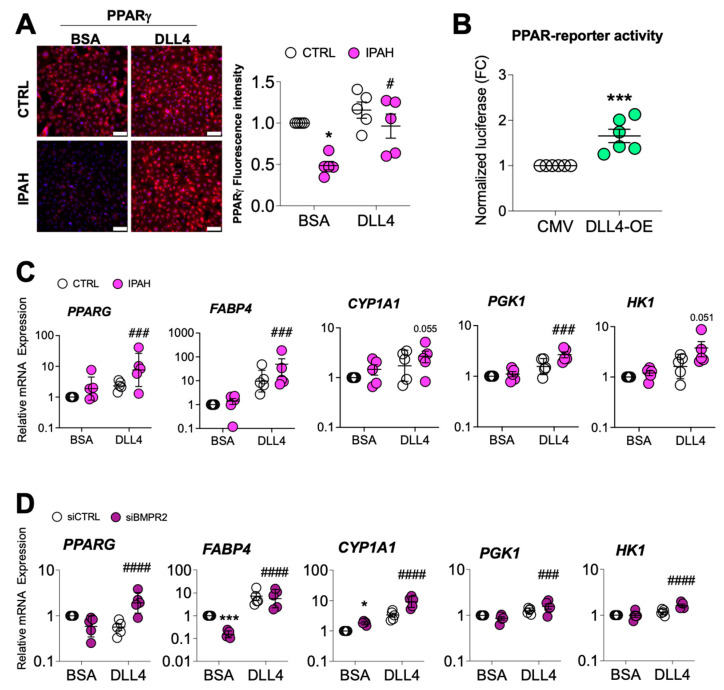
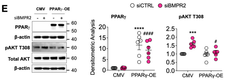
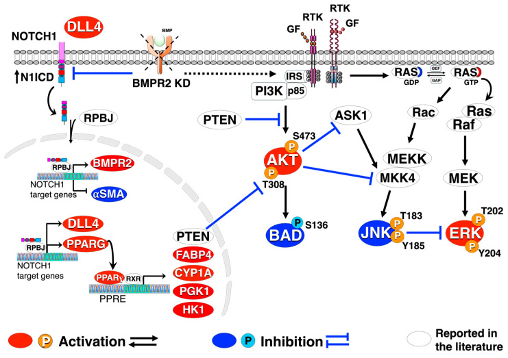
Pulmonary arterial hypertension (PAH) is a progressive cardiopulmonary disease characterized by pathologic vascular remodeling of small pulmonary arteries. Endothelial dysfunction in advanced PAH is associated with proliferation, apoptosis resistance, and endothelial to mesenchymal transition (EndoMT) due to aberrant signaling. DLL4, a cell membrane associated NOTCH ligand, plays a pivotal role maintaining vascular integrity. Inhibition of DLL4 has been associated with the development of pulmonary hypertension, but the mechanism is incompletely understood. Here we report that BMPR2 silencing in pulmonary artery endothelial cells (PAECs) activated AKT and suppressed the expression of DLL4. Consistent with these in vitro findings, increased AKT activation and reduced DLL4 expression was found in the small pulmonary arteries of patients with PAH. Increased NOTCH1 activation through exogenous DLL4 blocked AKT activation, decreased proliferation and reversed EndoMT. Exogenous and overexpression of DLL4 induced BMPR2 and PPRE promoter activity, and BMPR2 and PPARG mRNA in idiopathic PAH (IPAH) ECs. PPARγ, a nuclear receptor associated with EC homeostasis, suppressed by BMPR2 loss was induced and activated by DLL4/NOTCH1 signaling in both BMPR2-silenced and IPAH ECs, reversing aberrant phenotypic changes, in part through AKT inhibition. Directly blocking AKT or restoring DLL4/NOTCH1/PPARγ signaling may be beneficial in preventing or reversing the pathologic vascular remodeling of PAH.

Keywords: AKT; BMPR2; DLL4; PPARγ; endothelial dysfunction; pulmonary arterial hypertension; signal transduction; vascular remodeling.


Conflict of interest statement

The authors declare no conflict of interest.

Figures

Figure 1
Figure 1
BMPR2 knockdown activates AKT and protects the cells against apoptosis. (A) Human primary pulmonary artery endothelial cells were transfected with control (siCTRL) or BMPR2 siRNA (siBMPR2) for 48 h and total protein lysates were collected. Western blots of BMPR2, phosphorylation of AKT at S473 and T308, total AKT, phosphorylation of BCL2-associated agonist of cell death (BAD) at S136 and total BAD were detected. Densitometric analysis of each protein relative to total β-actin, total AKT or total BAD and normalized to its corresponding siCTRL. Representative Western blots are shown, n = 5 independent experiments. (B,C) Immunofluorescence of pAKT (S473) (red), CD31 (green) antibodies and nuclear staining with Hoechst 33,342 (blue) in HPAH, IPAH and healthy control (CTRL) lung. Scale bar = 20 μm. Insert scale bar = 5 μm. (D) Caspase 3/7 activity was measured after 24 h withdrawal of serum and growth factors (S/GFW). Data presented as mean ± SEM; n = 5. (A) Paired t-test, * p < 0.05; ** p < 0.01; **** p < 0.001; (D) 2-way ANOVA with Tukey HSD ### p < 0.005 (siBMPR2-complete vs. siBMPR2- S/GFW).
Figure 2
Figure 2
Loss of activated JNK1 contributes to apoptosis resistance as measured by reduced caspase 3 cleavage and flow cytometry analysis. (A) Human primary pulmonary artery endothelial cells (PAECs) were transfected with control (siCTRL), BMPR2 (siBMPR2), or JNK1 (siJNK1) siRNA for 48 h and total protein lysates were collected and analyzed for phosphorylated JNK1 (pJNK), cleaved caspase 3, BIM and phosphorylated ERK (pERK). Densitometric analysis was performed relative to total JNK2, β-actin or total ERK and normalized to its corresponding siCTRL. Representative Western blots are shown (n = 5, 6 independent experiments). (B) After siRNA transfection (48 h), cells were cultured in either complete media or serum and growth factor-free media (S/GFW) for 24 h followed by staining with annexin and propidium iodide (PI) for 10 min and data acquired on MACSquant. (C) Column scatter plot representing the percentage of apoptotic cells (cells stained with annexin V not PI, quadrant 3) (n = 4). Data presented as mean ± SEM; (A) 1-way ANOVA with Tukey HSD, * p < 0.05; **** p < 0.001; (C) 2-way ANOVA with Tukey HSD; ## p < 0.01 (siCTRL- S/GFW vs. siBMPR2- S/GFW).
Figure 3
Figure 3
PI3Kδ inhibitor, leniolisib, decreases AKT activation, proliferation, and EndoMT while increasing apoptosis in BMPR2-silenced PAECs. (A) Human primary pulmonary artery endothelial cells (PAECs) were transfected with control (siCTRL) or BMPR2 (siBMPR2) siRNA for 48 h and then exposed to leniolisib for 4 h at the indicated concentrations (n = 5). (B) Effect of 10 μM leniolisib on phosphorylation of AKT (S473 and T308) in BMPR2- and CAV1-silenced PAECs. Representative Western blots are shown and densitometric analysis (mean ± SEM) relative to β-actin or total AKT and normalized to its corresponding siCTRL (n = 4, 5). (C) BrdU cell proliferation of PAECs transfected with control (siCTRL), BMPR2 (siBMPR2) or CAV1 (siCAV1) siRNA for 48 h then replated and treated with vehicle (DMSO), 1 μM or 10 μM leniolisib for an additional 72 h. (D) Leniolisib reactivated apoptosis as assessed by annexin and PI staining (n = 5). (E) Representative immunofluorescence of endothelial markers (VE-cadherin, vWF and CD31) and mesenchymal markers (α-SMA, SNAIL/SLUG and CD44) in PAECs transfected with control, BMPR2, or CAV1 siRNA for 48 h then treated with 10 μM leniolisib for 24 h (n = 5). Scale bar = 100 μm. Data presented as mean ± SEM. * p < 0.05 (siCTRL versus siBMPR2), # p < 0.05 (vehicle versus treatment).
Figure 3
Figure 3
PI3Kδ inhibitor, leniolisib, decreases AKT activation, proliferation, and EndoMT while increasing apoptosis in BMPR2-silenced PAECs. (A) Human primary pulmonary artery endothelial cells (PAECs) were transfected with control (siCTRL) or BMPR2 (siBMPR2) siRNA for 48 h and then exposed to leniolisib for 4 h at the indicated concentrations (n = 5). (B) Effect of 10 μM leniolisib on phosphorylation of AKT (S473 and T308) in BMPR2- and CAV1-silenced PAECs. Representative Western blots are shown and densitometric analysis (mean ± SEM) relative to β-actin or total AKT and normalized to its corresponding siCTRL (n = 4, 5). (C) BrdU cell proliferation of PAECs transfected with control (siCTRL), BMPR2 (siBMPR2) or CAV1 (siCAV1) siRNA for 48 h then replated and treated with vehicle (DMSO), 1 μM or 10 μM leniolisib for an additional 72 h. (D) Leniolisib reactivated apoptosis as assessed by annexin and PI staining (n = 5). (E) Representative immunofluorescence of endothelial markers (VE-cadherin, vWF and CD31) and mesenchymal markers (α-SMA, SNAIL/SLUG and CD44) in PAECs transfected with control, BMPR2, or CAV1 siRNA for 48 h then treated with 10 μM leniolisib for 24 h (n = 5). Scale bar = 100 μm. Data presented as mean ± SEM. * p < 0.05 (siCTRL versus siBMPR2), # p < 0.05 (vehicle versus treatment).
Figure 4
Figure 4
DLL4 is decreased in BMPR2-silenced PAECs and in lung tissue from patients with pulmonary arterial hypertension (PAH). Human primary pulmonary artery endothelial cells (PAECs) were transfected with control (siCTRL) or BMPR2 (siBMPR2) siRNA for 48 h and total protein lysates were collected for Western blotting of (A) DLL4, (B) N1ICD, (C) NOTCH1, (D) N2ICD, and (E) N4ICD. (F) DLL4 protein was also analyzed in PAECs transfected with either control (siCTRL) or BMPR2 (siBMPR2), CAV1 (siCAV1) or SMAD9 (siSMAD9) gene-specific siRNA pools. Representative Western blots are shown and densitometric analysis relative to β-actin and normalized to its corresponding siCTRL (n = 5). (G) Immunohistochemical staining of DLL4 in paraffin embedded lung of failed donor controls (CTRL; n = 5), HPAH (n = 3) and IPAH (n = 2). Scale bar = 50 μm. Insert scale bar = 20 μm Data are presented as mean ± SEM; n = 5. (AE) paired t-test, (F) 1-way ANOVA with Tukey HSD * p < 0.05; *** p < 0.005; and **** p < 0.001.
Figure 5
Figure 5
Immobilized DLL4 activates NOTCH1 signaling inhibiting AKT activation, proliferation and EndoMT. Human primary pulmonary artery endothelial cells (PAECs) were grown on BSA- or DLL4-coated plates and the following day the cells were transfected with either non-targeting control (siCTRL) or BMPR2 (siBMPR2) gene-specific siRNA pools. After BMPR2 knockdown (48 h), total protein lysates were collected and analyzed by Western blotting for expression of (A) DLL4, N1ICD, NOTCH1, N2ICD and N4ICD or (B) phosphorylated AKT (pAKT S473 and pAKT T308) or phosphorylated ERK (pERK). Representative Western blots are shown (n = 4–6, as indicated). Densitometric analysis relative to β-actin, total AKT or total ERK and normalized to its corresponding siCTRL. (C) BrdU cell proliferation of PAECs transfected with control or BMPR2 siRNA for 48 h and then replated onto BSA- or DLL4-coated plates for an additional 48 h (n = 5). (D) BrdU cell proliferation of healthy and IPAH ECs were grown on either BSA or DLL4 for 48 h (n = 5). (E,F) Immunofluorescence staining of α-SMA (ACTA2) and VE-Cadherin (CDH5) in ECs from healthy and IPAH grown on either BSA or DLL4. Scale bar = 100 μm. Data are presented as mean ± SEM, 2-way ANOVA with Tukey HSD; * p < 0.05; ** p < 0.01; **** p < 0.001 (siCTRL-BSA versus siBMPR2-BSA); # p < 0.05; ### p < 0.005; and #### p < 0.001 (siBMPR2-BSA versus siBMPR2-DLL4).
Figure 6
Figure 6
DLL4 increases BMPR2 transcription. (A,B) Quantitative RT-PCR of BMPR2 mRNA from (A) PAECs or from (B) healthy or IPAH PAECs grown on BSA- or DLL4-coated plates. (C) Schematic of BMPR2 promoter with putative RBPJ (recombination signal binding protein for immunoglobulin kappa J region) binding sites indicated. (D) BMPR2 promoter activity in PAECs grown on BSA- or DLL4-coated plates and transfected with the promoter driven luciferase reporter. (E) BMPR2 promoter activity in PAECs transfected with the promoter driven luciferase reporter plus either empty vector control (CMV) or DLL4 overexpression plasmid (DLL4-OE). For quantitative RT-PCR, data are presented as the geometric mean ± SD; n = 4, 5. For promoter activity, data presented as mean ± SEM; n = 6. Paired t-test, * p < 0.05; **** p < 0.001; and ## p < 0.01 (siBMPR2-BSA versus siBMPR2-DLL4).
Figure 7
Figure 7
DLL4-induced NOTCH1 activation rescues PPARγ expression. (A) PPARγ immunofluorescence staining of PAECs endothelial cells from healthy or IPAH patients grown on either BSA or DLL4 (n = 5). Scale bar = 100 μm. (B) Human primary pulmonary artery endothelial cells (PAECs) were transfected with empty vector (CMV) or DLL4 overexpression plasmid (DLL4-OE) for 24 h and PPAR-driven reporter activity assessed (n = 6). (C) mRNA levels from healthy (CTRL) or IPAH ECs grown on BSA or DLL4 for 48 h were analyzed for PPARG expression and PPARγ target genes. (D) mRNA from PAECs transfected with control (siCTRL) or BMPR2 (siBMPR2) siRNA and grown on BSA or DLL4 for 48 h were analyzed for PPARG expression and PPARγ target genes. (E) After 24 h of gene silencing with either control or BMPR2 siRNAs, PAECs were then transfected with empty vector control (CMV) or PPARγ overexpression plasmid for 24 h and expression of PPARγ and phosphorylated AKT (pAKT T308) protein was analyzed by Western blotting. Densitometric quantification relative to β-actin or total AKT and normalized to its corresponding siCTRL. Representative Western blots are shown. Data presented as mean ± SEM; n = 6. The mRNA levels were measured by quantitative RT-PCR and presented as the geometric mean ± SD; n = 5. 2-way ANOVA with Tukey HSD; * p < 0.05; *** p < 0.005 (siCTRL-BSA versus siBMPR2-BSA); # p < 0.05; ### p < 0.005; and #### p < 0.001 (siBMPR2-BSA versus siBMPR2-DLL4).
Figure 7
Figure 7
DLL4-induced NOTCH1 activation rescues PPARγ expression. (A) PPARγ immunofluorescence staining of PAECs endothelial cells from healthy or IPAH patients grown on either BSA or DLL4 (n = 5). Scale bar = 100 μm. (B) Human primary pulmonary artery endothelial cells (PAECs) were transfected with empty vector (CMV) or DLL4 overexpression plasmid (DLL4-OE) for 24 h and PPAR-driven reporter activity assessed (n = 6). (C) mRNA levels from healthy (CTRL) or IPAH ECs grown on BSA or DLL4 for 48 h were analyzed for PPARG expression and PPARγ target genes. (D) mRNA from PAECs transfected with control (siCTRL) or BMPR2 (siBMPR2) siRNA and grown on BSA or DLL4 for 48 h were analyzed for PPARG expression and PPARγ target genes. (E) After 24 h of gene silencing with either control or BMPR2 siRNAs, PAECs were then transfected with empty vector control (CMV) or PPARγ overexpression plasmid for 24 h and expression of PPARγ and phosphorylated AKT (pAKT T308) protein was analyzed by Western blotting. Densitometric quantification relative to β-actin or total AKT and normalized to its corresponding siCTRL. Representative Western blots are shown. Data presented as mean ± SEM; n = 6. The mRNA levels were measured by quantitative RT-PCR and presented as the geometric mean ± SD; n = 5. 2-way ANOVA with Tukey HSD; * p < 0.05; *** p < 0.005 (siCTRL-BSA versus siBMPR2-BSA); # p < 0.05; ### p < 0.005; and #### p < 0.001 (siBMPR2-BSA versus siBMPR2-DLL4).
Figure 8
Figure 8
Reactivation of DLL4/NOTCH1 signaling blocks AKT activation, inhibiting proliferation and endothelial-to-mesenchymal transition. Crosstalk among DLL4/NOTCH1, BMPR2, PPARɣ, and AKT signaling pathways. BMPR2 knockdown (KD) culminates in sustained AKT and ERK activation along with JNK1 inactivation leading to an apoptosis-resistant, hyper-proliferative, endothelial-to-mesenchymal transition (EndoMT). Loss of BMPR2 also results in decreased DLL4/N1ICD and PPARɣ protein. Activation of DLL4/N1ICD signaling increases BMPR2/PPARɣ expression blocking AKT activation leading to a decrease in proliferation and EndoMT.

Update of

References

    1. Archer S., Rich S. Primary pulmonary hypertension: A vascular biology and translational research “Work in progress”. Circulation. 2000;102:2781–2791. doi: 10.1161/01.CIR.102.22.2781. - DOI - PubMed
    1. Bisserier M., Pradhan N., Hadri L. Current and emerging therapeutic approaches to pulmonary hypertension. Rev. Cardiovasc. Med. 2020;21:163–179. - PMC - PubMed
    1. Wilken E., Bennji S., Symons G., Williams P.G., Allwood B. Treatment of pulmonary arterial hypertension: A review of drugs available for advanced therapy. Afr. J. Thorac. Crit. Care Med. 2019;25:14–21. doi: 10.7196/SARJ.2019.v25i1.236. - DOI - PMC - PubMed
    1. Sakao S., Taraseviciene-Stewart L., Lee J.D., Wood K., Cool C.D., Voelkel N.F. Initial apoptosis is followed by increased proliferation of apoptosis-resistant endothelial cells. FASEB J. 2005;19:1178–1180. doi: 10.1096/fj.04-3261fje. - DOI - PubMed
    1. Tang H., Chen J., Fraidenburg D.R., Song S., Sysol J.R., Drennan A.R., Offermanns S., Ye R.D., Bonini M.G., Minshall R.D., et al. Deficiency of Akt1, but not Akt2, attenuates the development of pulmonary hypertension. Am. J. Physiol. Lung Cell Mol. Physiol. 2015;308:L208–L220. doi: 10.1152/ajplung.00242.2014. - DOI - PMC - PubMed

MeSH terms

LinkOut - more resources