. 2024 May 20;16(10):1945.
doi: 10.3390/cancers16101945.

Immunohistochemical Investigation into Protein Expression Patterns of FOXO4, IRF8 and LEF1 in Canine Osteosarcoma

Affiliations

Immunohistochemical Investigation into Protein Expression Patterns of FOXO4, IRF8 and LEF1 in Canine Osteosarcoma

Simone de Brot et al. Cancers (Basel). .

Abstract

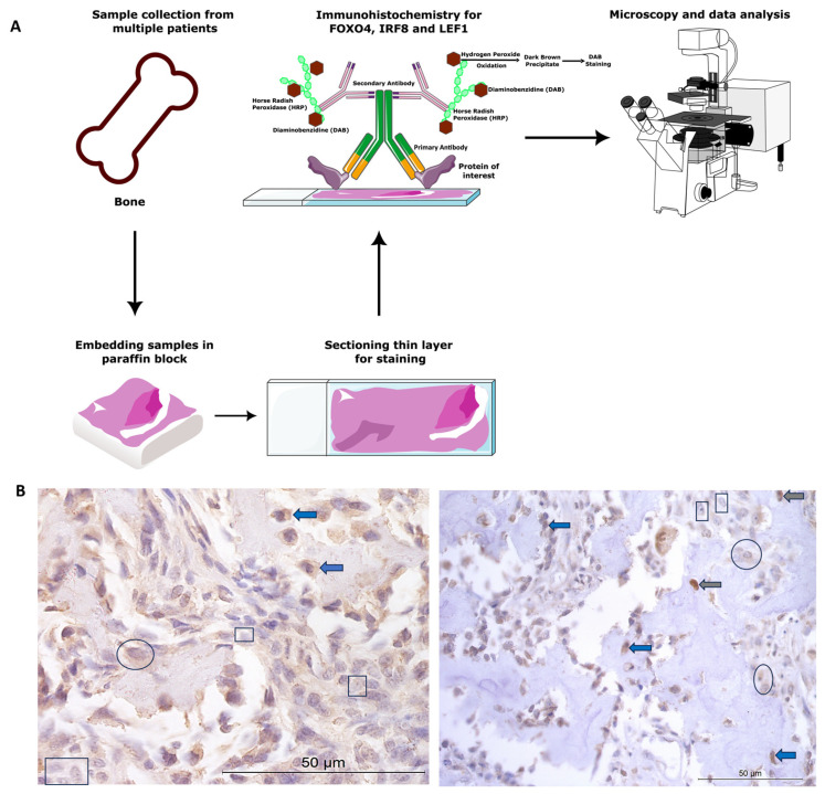
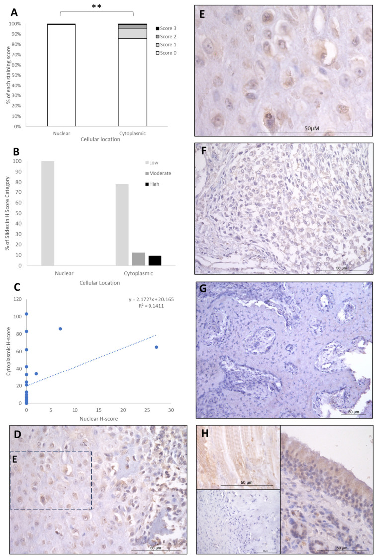
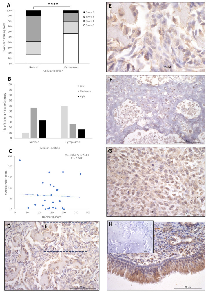
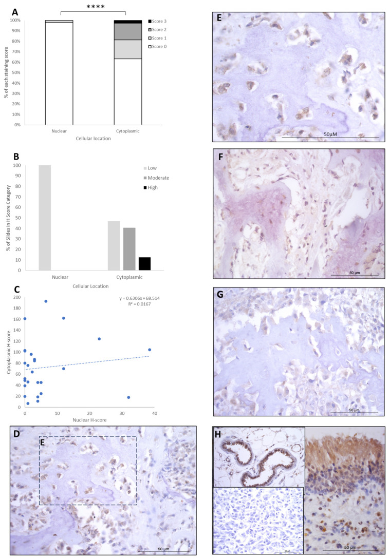
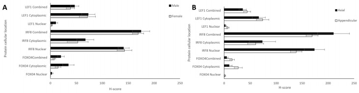
Osteosarcoma (OSA) is the most common type of primary bone malignancy in people and dogs. Our previous molecular comparisons of canine OSA against healthy bone resulted in the identification of differentially expressed protein-expressing genes (forkhead box protein O4 (FOXO4), interferon regulatory factor 8 (IRF8), and lymphoid enhancer binding factor 1 (LEF1)). Immunohistochemistry (IHC) and H-scoring provided semi-quantitative assessment of nuclear and cytoplasmic staining alongside qualitative data to contextualise staining (n = 26 patients). FOXO4 was expressed predominantly in the cytoplasm with significantly lower nuclear H-scores. IRF8 H-scores ranged from 0 to 3 throughout the cohort in the nucleus and cytoplasm. LEF1 was expressed in all patients with significantly lower cytoplasmic staining compared to nuclear. No sex or anatomical location differences were observed. While reduced levels of FOXO4 might indicate malignancy, the weak or absent protein expression limits its primary use as diagnostic tumour marker. IRF8 and LEF1 have more potential for prognostic and diagnostic uses and facilitate further understanding of their roles within their respective molecular pathways, including Wnt/beta-catenin/LEF1 signalling and differential regulation of tumour suppressor genes. Deeper understanding of the mechanisms involved in OSA are essential contributions towards the development of novel diagnostic, prognostic, and treatment options in human and veterinary medicine contexts.

Keywords: cancer identification; forkhead box protein O4; interferon regulatory factor 8; lymphoid enhancer binding factor 1; osteosarcoma; pathology.


Conflict of interest statement

The authors declare no conflicts of interest.

Figures

Figure 1
Figure 1
Canine osteosarcoma protein expression materials and methods. (A) Overview of methods. (B) Examples of cytoplasmic and nuclear H-scores 0, 1, 2, and 3, on IRF8 and LEF1 immunohistochemistry photomicrographs. H-score 0 (rectangles), 1+ (circles), 2+ (blue arrows), 3+ (grey arrows).
Figure 2
Figure 2
FOXO4 cytoplasmic and nuclear H-scores. (A) H-score (scores 0, 1, 2, and 3) distributions. (B) H-score low/moderate/high classifications across the cases (p = 0.02). (C) Nuclear and cytoplasmic H-score distributions and correlation. Overall, the nuclear H-scores were significantly lower than cytoplasmic (** p = 0.002), n = 26. (DG) Immunohistochemical staining photomicrographs of canine osteosarcoma FOXO4 expression, 40× magnification. (H) Right-hand side: positive control nasal mucosa lined by well-differentiated pseudostratified tall columnar ciliated epithelium, inset upper left: muscle positive control, insert lower left: negative control, 40× magnification. All scale bars represent 50 µm.
Figure 3
Figure 3
IRF8 cytoplasmic and nuclear H-scores. (A) H-score (scores 0, 1, 2, and 3) distributions. (B) H-score low/moderate/high classifications across the cases (p = 0.0001). (C) Nuclear and cytoplasmic H-score distributions and correlation. Overall, the nuclear scores were significantly higher than cytoplasmic (**** p = 0.0001), n = 26. (DG) Immunohistochemical staining photomicrographs of canine osteosarcoma IRF8 expression, 40× magnification. (H) Positive control nasal mucosa lined by well-differentiated pseudostratified tall columnar ciliated epithelium, inset upper left: negative control, 40× magnification. All scale bars represent 50 µm.
Figure 4
Figure 4
LEF1 cytoplasmic and nuclear H-scores. (A) H-score (scores 0, 1, 2, and 3) distributions. (B) H-score low/moderate/high classifications across the cases (p = 0.0001). (C) Nuclear and cytoplasmic H-score distributions and correlation. Overall, the nuclear scores were significantly lower than cytoplasmic (**** p = 0.0001), n = 26. (DG) Immunohistochemical staining photomicrographs of canine osteosarcoma LEF1 expression, 40× magnification. (H) Right-hand side: positive control nasal mucosa lined by well-differentiated pseudostratified tall columnar ciliated epithelium, inset upper left: endothelial cells of vasculature positive control, inset lower left: negative control, 40× magnification. All scale bars represent 50 µm.
Figure 5
Figure 5
Nuclear, cytoplasmic, and combined H-scores for FOXO4, IRF8, and LEF1 by sex and anatomical location. (A) Males and females (n = 12 and 13, respectively), and (B) differing bone locations—appendicular and axial (n = 20 and 6, respectively). No statistically significant differences in sex or bone location were observed for nuclear, cytoplasmic, or total H-scores (t-test, p > 0.05).

Similar articles

Cited by

  • Comparison of Differentially Expressed Genes in Human and Canine Osteosarcoma.
    Jackson-Oxley J, Alibhai AA, Guerin J, Thompson R, Patke R, Harris AE, Woodcock CL, Varun D, Haque M, Modikoane TK, Kumari AA, Lothion-Roy J, Brot S, Dunning MD, Jeyapalan JN, Mongan NP, Rutland CS. Jackson-Oxley J, et al. Life (Basel). 2025 Jun 12;15(6):951. doi: 10.3390/life15060951. Life (Basel). 2025. PMID: 40566602 Free PMC article.

References

    1. Schmidt A.F., Groenwold R.H.H., Amsellem P., Bacon N., Klungel O.H., Hoes A.W., de Boer A., Kow K., Maritato K., Kirpensteijn J., et al. Which dogs with appendicular osteosarcoma benefit most from chemotherapy after surgery? Results from an individual patient data meta-analysis. Prev. Vet. Med. 2016;125:116–125. doi: 10.1016/j.prevetmed.2015.10.016. - DOI - PubMed
    1. Cook M.R., Lorbach J., Husbands B.D., Kisseberth W.C., Samuels S., Silveira C., Wustefeld-Janssens B.G., Wouda R., Keepman S., Oblak M.L., et al. A retrospective analysis of 11 dogs with surface osteosarcoma. Vet. Comp. Oncol. 2022;20:82–90. doi: 10.1111/vco.12741. - DOI - PubMed
    1. Greene S. Withrow & MacEwen’s Small Animal Clinical Oncology, 6th edition. Am. J. Vet. Res. 2020;81:391.
    1. Morello E., Martano M., Buracco P. Biology, diagnosis and treatment of canine appendicular osteosarcoma: Similarities and differences with human osteosarcoma. Vet. J. 2011;189:268–277. doi: 10.1016/j.tvjl.2010.08.014. - DOI - PubMed
    1. Selvarajah G.T., Kirpensteijn J. Prognostic and predictive biomarkers of canine osteosarcoma. Vet. J. 2010;185:28–35. doi: 10.1016/j.tvjl.2010.04.010. - DOI - PubMed