

The Role of POCUS to Face COVID-19: A Narrative Review
- PMID: 38792298
- PMCID: PMC11121862
- DOI: 10.3390/jcm13102756
The Role of POCUS to Face COVID-19: A Narrative Review
Abstract
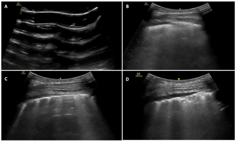
COVID-19 has been a challenging outbreak to face, with millions of deaths among the globe. Acute respiratory failure due to interstitial pneumonia was the leading cause of death other than prothrombotic activation and complications. Lung ultrasound (LUS) and point-of-care ultrasound (POCUS) are widely used not only to triage, to identify, and to monitor lungs involvement but also to assess hemodynamic status and thrombotic and hemorrhagic complications, mainly in critically ill patients. POCUS has gained growing consideration due to its bedside utilization, reliability, and reproducibility even in emergency settings especially in unstable patients. In this narrative review, we aim to describe LUS and POCUS utilization in COVID-19 infection based on the literature found on this topic. We reported the LUS patterns of COVID-19 pulmonary infection, the diagnostic accuracy with respect to CT lung scan, its prognostic value, the variety of scores and protocols proposed, and the utilization of POCUS to investigate the extra-lung complications.
Keywords: COVID-19; acute heart failure; acute respiratory failure; deep venous thrombosis; diaphragm impairment; lung effusion; lung ultrasound; pneumonia; pulmonary embolism.
Conflict of interest statement
The authors declare no conflicts of interest.
Figures
References
-
- Bellani G., Laffey J.G., Pham T., Fan E., Brochard L., Esteban A., Gattinoni L., Van Haren F., Larsson A., McAuley D.F., et al. Epidemiology, patterns of care, and mortality for patients with acute respiratory distress syndrome in intensive care units in 50 countries. JAMA. 2016;315:788–800. doi: 10.1001/jama.2016.0291. - DOI - PubMed
Publication types
LinkOut - more resources
Full Text Sources
