Review
. 2024 May 17;13(10):2966.
doi: 10.3390/jcm13102966.

Intraoperative Neurophysiological Monitoring in Neurosurgery

Affiliations
Review

Intraoperative Neurophysiological Monitoring in Neurosurgery

Giusy Guzzi et al. J Clin Med. .

Abstract

Intraoperative neurophysiological monitoring (IONM) is a crucial advancement in neurosurgery, enhancing procedural safety and precision. This technique involves continuous real-time assessment of neurophysiological signals, aiding surgeons in timely interventions to protect neural structures. In addition to inherent limitations, IONM necessitates a detailed anesthetic plan for accurate signal recording. Given the growing importance of IONM in neurosurgery, we conducted a narrative review including the most relevant studies about the modalities and their application in different fields of neurosurgery. In particular, this review provides insights for all physicians and healthcare professionals unfamiliar with IONM, elucidating commonly used techniques in neurosurgery. In particular, it discusses the roles of IONM in various neurosurgical settings such as tumoral brain resection, neurovascular surgery, epilepsy surgery, spinal surgery, and peripheral nerve surgery. Furthermore, it offers an overview of the anesthesiologic strategies and limitations of techniques essential for the effective implementation of IONM.

Keywords: direct cortical stimulation; electrocorticography; electromyography; evoked potentials; intraoperative neurophysiological monitoring; neurosurgery.


Conflict of interest statement

Federico Longhini contributed to the development of a new helmet for mechanical ventilation and he is designated as the inventor (European Patent number 3320941) not related to the present manuscript. He also received speaking fees from Draeger, Intersurgical, and Fisher & Paykel. The remaining authors have no relevant financial or non-financial interests to disclose.

Figures
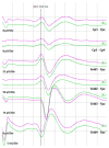
Figure 1
Figure 1
Phase reversal achieved by stimulating the right median nerve and recording at the cortical level with a subdural strip containing 4 contacts. The technique was used to identify the Rolandic sulcus, which in this case is identified by the phase reversal between contacts 3 and 4 of the strip.
Figure 2
Figure 2
SSEPs of the upper limbs with stimulation from the median nerve using adhesive electrodes and recording from the scalp with corkscrew electrodes positioned according to the International 10–20 system.
Figure 3
Figure 3
Transcranial motor evoked potentials (MEP) obtained by stimulating with corkscrew electrodes in the C3–FZ position at a threshold of 90 mA in a case of left parietal lesion.
Figure 4
Figure 4
Direct cortical stimulation. Motor evoked potentials (MEP) obtained by stimulating from contacts on a cortical strip, positioned at the cortical level, with reference to a corkscrew electrode placed on the scalp. Panels from left to right show the responses obtained from the biceps muscle, the ulnar extensor of the wrist, the abductor of the fifth finger, and the short abductor of the thumb.
Figure 5
Figure 5
Subcortical stimulation. In a left parietal expansive lesion, during the removal phase, subcortical stimulation was performed with a suction probe, referenced to a corkscrew electrode placed on the scalp. Activation of the corticospinal tract was recorded at an intensity of 5.5 mA.
Figure 6
Figure 6
Motor evoked potentials in spinal surgery. The figure reports the MEP obtained, from top to bottom, in the left and right iliopsoas, vastus lateralis, tibialis anterior, abductor hallucis, gastrocnemius, and anal sphincter in a case of cauda surgery. The stimulation was obtained with a bipolar probe at 0.4 mA.

References

    1. Grosland J.O., Todd M.M., Goldstein P.A. Neuromonitoring in the ambulatory anesthesia setting: A pro-con discussion. Curr. Opin. Anaesthesiol. 2018;31:667–672. doi: 10.1097/ACO.0000000000000654. - DOI - PubMed
    1. Zebian B., Vergani F., Lavrador J.P., Mukherjee S., Kitchen W.J., Stagno V., Chamilos C., Pettorini B., Mallucci C. Recent technological advances in pediatric brain tumor surgery. CNS Oncol. 2017;6:71–82. doi: 10.2217/cns-2016-0022. - DOI - PMC - PubMed
    1. Lall R.R., Hauptman J.S., Munoz C., Cybulski G.R., Koski T., Ganju A., Fessler R.G., Smith Z.A. Intraoperative neurophysiological monitoring in spine surgery: Indications, efficacy, and role of the preoperative checklist. Neurosurg. Focus. 2012;33:E10. doi: 10.3171/2012.9.FOCUS12235. - DOI - PubMed
    1. Moehl K., Shandal V., Anetakis K., Paras S., Mina A., Crammond D., Balzer J., Thirumala P.D. Predicting transient ischemic attack after carotid endarterectomy: The role of intraoperative neurophysiological monitoring. Clin. Neurophysiol. 2022;141:1–8. doi: 10.1016/j.clinph.2022.06.010. - DOI - PubMed
    1. Szelenyi A., Fernandez-Conejero I., Kodama K. Surgery and intraoperative neurophysiologic monitoring for aneurysm clipping. Handb. Clin. Neurol. 2022;186:375–393. - PubMed

LinkOut - more resources