

Characterization Methods for Nanoparticle-Skin Interactions: An Overview
- PMID: 38792620
- PMCID: PMC11122446
- DOI: 10.3390/life14050599
Characterization Methods for Nanoparticle-Skin Interactions: An Overview
Abstract
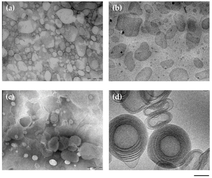
Research progresses have led to the development of different kinds of nanoplatforms to deliver drugs through different biological membranes. Particularly, nanocarriers represent a precious means to treat skin pathologies, due to their capability to solubilize lipophilic and hydrophilic drugs, to control their release, and to promote their permeation through the stratum corneum barrier. A crucial point in the development of nano-delivery systems relies on their characterization, as well as in the assessment of their interaction with tissues, in order to predict their fate under in vivo administration. The size of nanoparticles, their shape, and the type of matrix can influence their biodistribution inside the skin strata and their cellular uptake. In this respect, an overview of some characterization methods employed to investigate nanoparticles intended for topical administration is presented here, namely dynamic light scattering, zeta potential, scanning and transmission electron microscopy, X-ray diffraction, atomic force microscopy, Fourier transform infrared and Raman spectroscopy. In addition, the main fluorescence methods employed to detect the in vitro nanoparticles interaction with skin cell lines, such as fluorescence-activated cell sorting or confocal imaging, are described, considering different examples of applications. Finally, recent studies on the techniques employed to determine the nanoparticle presence in the skin by ex vivo and in vivo models are reported.
Keywords: confocal microscopy; fluorescence microscopy; hyperspectral microscopy; nanoparticles; skin; transmission electron microscopy.
Conflict of interest statement
The authors declare no conflicts of interest.
Figures


References
-
- Bielfeldt S., Bonnier F., Byrne H.J., Chourpa I., Dancik Y., Lane M.E., Lunter D.J., Munnier E., Puppels G., Tfayli A., et al. Monitoring Dermal Penetration and Permeation Kinetics of Topical Products; the Role of Raman Microspectroscopy. TrAC Trends Anal. Chem. 2022;156:116709. doi: 10.1016/J.TRAC.2022.116709. - DOI
-
- Hallan S.S., Sguizzato M., Drechsler M., Mariani P., Montesi L., Cortesi R., Björklund S., Ruzgas T., Esposito E. The Potential of Caffeic Acid Lipid Nanoparticulate Systems for Skin Application: In Vitro Assays to Assess Delivery and Antioxidant Effect. Nanomaterials. 2021;11:171. doi: 10.3390/NANO11010171. - DOI - PMC - PubMed
Publication types
Grants and funding
LinkOut - more resources
Full Text Sources
