Review
. 2024 May 16;12(5):1004.
doi: 10.3390/microorganisms12051004.

Exploring the Toxin-Mediated Mechanisms in Clostridioides difficile Infection

Affiliations
Review

Exploring the Toxin-Mediated Mechanisms in Clostridioides difficile Infection

Evdokia Pourliotopoulou et al. Microorganisms. .

Abstract

Clostridioides difficile infection (CDI) is the leading cause of nosocomial antibiotic-associated diarrhea, and colitis, with increasing incidence and healthcare costs. Its pathogenesis is primarily driven by toxins produced by the bacterium C. difficile, Toxin A (TcdA) and Toxin B (TcdB). Certain strains produce an additional toxin, the C. difficile transferase (CDT), which further enhances the virulence and pathogenicity of C. difficile. These toxins disrupt colonic epithelial barrier integrity, and induce inflammation and cellular damage, leading to CDI symptoms. Significant progress has been made in the past decade in elucidating the molecular mechanisms of TcdA, TcdB, and CDT, which provide insights into the management of CDI and the future development of novel treatment strategies based on anti-toxin therapies. While antibiotics are common treatments, high recurrence rates necessitate alternative therapies. Bezlotoxumab, targeting TcdB, is the only available anti-toxin, yet limitations persist, prompting ongoing research. This review highlights the current knowledge of the structure and mechanism of action of C. difficile toxins and their role in disease. By comprehensively describing the toxin-mediated mechanisms, this review provides insights for the future development of novel treatment strategies and the management of CDI.

Keywords: Clostridioides difficile; actin cytoskeleton; bacterial toxins; infection; inflammation; pathogenesis.


Conflict of interest statement

The authors declare no conflicts of interest.

Figures

Figure 1
Figure 1
(A) Pathogenicity Locus (PaLoc). The genes tcdA and tcdB (pink arrows) encode the toxins TcdA and TcdB, respectively. The regulatory genes tcdR (positive) and tcdC (negative) modulate the transcription of tcdA and tcdB and are presented with green arrows. Genes tcdE and tcdL (grey arrows) encode a holin and an endolysin, respectively, which are involved in toxin secretion. The direction of the arrows represents the direction of transcription of the genes. (B) TcdA and TcdB are divided into four domains: the glycosyltransferase domain (GTD; red), the autoprotease domain (APD; blue), the delivery and receptor-binding domain (DRBD; yellow), and the combined repetitive oligopeptides (CROPs; green).
Figure 2
Figure 2
Mechanism of action of TcdA and TcdB. Toxins bind surface receptors on the colonic epithelium and are endocytosed in acidic endosomes. Low pH triggers a conformational change in the toxins resulting in pore formation and translocation of GTD and APD in the cytosol. The activation of APD results in the cleavage and release of the GTD. The GTD blocks the function of Rho and Ras GTPases by transferring the UDP-glycose to GTPases, resulting in the induction of cytoskeletal damage.
Figure 3
Figure 3
Representation of CDI-mediated inflammatory host response. Once TcdA and TcdB destroy the intestinal epithelium, they cause damage to deeper layers of tissue such as the destruction of the myofibroblasts. The presence of toxins triggers the release of dendritic cells, neutrophils, monocytes, and macrophages from the blood vessels. IL-1β and IL-8 produced by the intestinal epithelial cells enhance inflammation and attract neutrophils to the lumen of the colon. Within, neutrophils form pseudomembranes. At the same time, toxins induce the degranulation of mast cells and the release of substance P (SP) and Calcitonin gene-related peptide (CGRP) from neurons of the enteric nervous system.
Figure 4
Figure 4
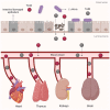
During CDI, C. difficile toxins TcdA and TcdB can breach the intestinal barrier and enter the bloodstream, resulting in systemic toxemia. Elevated levels of these toxins in the bloodstream can cause damage outside the colon, leading to dysfunction in multiple organs such as the heart, thymus, kidneys, and brain.
Figure 5
Figure 5
(A) Representation of CDT locus (CdtLoc). Genes cdtA and cdtB (grey arrows) encode the components CDTa and CDTb, respectively. The transcription of cdtA and cdtB is positively regulated by the regulatory gene cdtR (green arrow). The direction of the arrows represents the direction of transcription of the genes. (B) Schematic representation of CDTa and CDTb components of CDT. CDTa is divided into two regions: the N-terminal region and the C-terminal region. CDTb is divided into four conserved functional regions (Regions I–IV).
Figure 6
Figure 6
Schematic representation of CDT binding to cellular receptor and entry into the cell. CDTb binds into LSR and oligomerizes on the cell surface. Therefore, CDTa binds to the oligomeric form, and the complex is internalized into cells. The acidic environment of endosomes triggers conformational changes in CDTb resulting in a pore formation in the endosomal membrane and the translocation of CDTa into the cytosol. CDTa catalyzes the ADP-ribosylation of actin resulting in the disruption of F-actin and the formation of microtubule protrusions.

References

    1. Burke K.E., Lamont J.T. Clostridium difficile infection: A worldwide disease. Gut Liver. 2014;8:1–6. doi: 10.5009/gnl.2014.8.1.1. - DOI - PMC - PubMed
    1. Smits W.K., Lyras D., Lacy D.B., Wilcox M.H., Kuijper E.J. Clostridium difficile infection. Nat. Rev. Dis. Primers. 2016;2:16020. doi: 10.1038/nrdp.2016.20. - DOI - PMC - PubMed
    1. Warny M., Pepin J., Fang A., Killgore G., Thompson A., Brazier J., Frost E., McDonald L.C. Toxin production by an emerging strain of Clostridium difficile associated with outbreaks of severe disease in North America and Europe. Lancet. 2005;366:1079–1084. doi: 10.1016/s0140-6736(05)67420-x. - DOI - PubMed
    1. Geric B., Rupnik M., Gerding D.N., Grabnar M., Johnson S. Distribution of Clostridium difficile variant toxinotypes and strains with binary toxin genes among clinical isolates in an American hospital. J. Med. Microbiol. 2004;53:887–894. doi: 10.1099/jmm.0.45610-0. - DOI - PubMed
    1. Carter G.P., Rood J.I., Lyras D. The role of toxin A and toxin B in the virulence of Clostridium difficile. Trends Microbiol. 2012;20:21–29. doi: 10.1016/j.tim.2011.11.003. - DOI - PubMed

LinkOut - more resources