

Laser Forced Dehydration of Benign Vascular Lesions of the Oral Cavity: A Valid Alternative to Surgical Techniques
- PMID: 38793005
- PMCID: PMC11122876
- DOI: 10.3390/medicina60050822
Laser Forced Dehydration of Benign Vascular Lesions of the Oral Cavity: A Valid Alternative to Surgical Techniques
Abstract
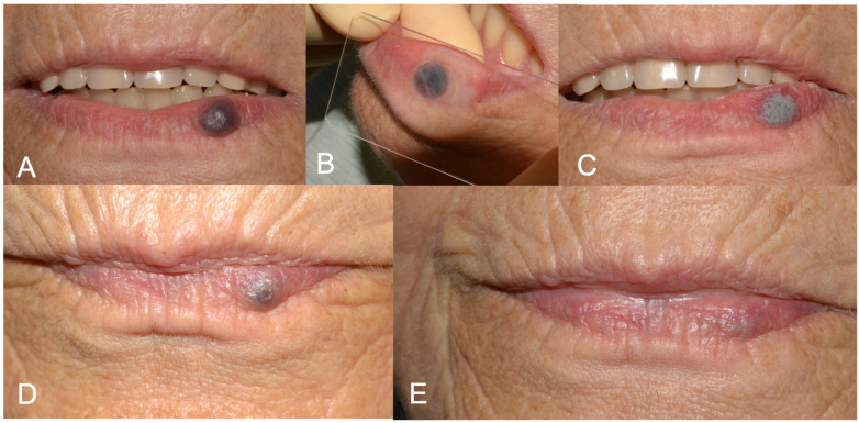
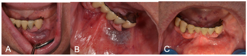
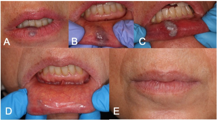
Background and Objectives: Low-flow vascular lesions are commonly encountered in the oral cavity and may require removal due to aesthetic concerns, repeated bleeding or a cluttering sensation. Laser devices represent an excellent aid due to their affinity with blood and to their biostimulating properties and have been substituting traditional excision in selected cases. Materials and Methods: In this study, 30 patients presenting with low-flow oral vascular lesions were included. The lesions were clinically evaluated as follows: lesion's site, reason for treatment, lesion's dimensions, confirmation of positive diascopy via compression with a glass slide and photograph. The lesions were treated with laser forced dehydration (LFD) and then followed-up after 3 weeks, 6 months and 1 year. The laser source was a K-Laser Blu Derma (Eltech, K-Laser S.r.l., Via Castagnole, 20/H, Treviso, Italy). In the case of incomplete healing, a further protocol was performed at the three-week follow-up, and a further follow-up was scheduled for three weeks after. The following aspects were evaluated at each appointment: pain, using a Numeric Rating Scale (NRS) from 0 to 10 (0 = no pain, 10 = worst pain ever); the need to take painkillers (day of intervention and during follow-up); bleeding (yes/no); scar formation. Results: Complete regression was obtained in all patients, with no side effects. Only one patient required a second LFD protocol. NRS was 0 for all patients for the whole duration of the follow-up. None of the patients took painkillers on the day of the intervention and during the follow-up. One patient declared slight bleeding the day of the intervention, which she easily managed at home. One patient showed a small non-retracting and non-painful scar at the three-week follow-up. No recurrences were found after six months and one year. Conclusions: LFD targets endogenous chromophores, minimizing damage to adjacent tissue and limiting side effects. LFD is effective and could be considered a conservative alternative to traditional excision in low-flow lesions.
Keywords: angioma; blue light; conservative; diode laser; vascular malformation.
Conflict of interest statement
The authors declare no conflicts of interest.
Figures
Similar articles
-
Blue light diode laser for treating benign maxillofacial vascular lesions: comparison of various techniques using the same diode laser.J Cosmet Laser Ther. 2024 Nov 16;26(5-8):122-128. doi: 10.1080/14764172.2024.2433215. Epub 2024 Nov 28. J Cosmet Laser Ther. 2024. PMID: 39606942
-
Blue diode laser versus traditional infrared diode laser and quantic molecular resonance scalpel: clinical and histological findings after excisional biopsy of benign oral lesions.J Biomed Opt. 2017 Dec 1;22(12):121602. doi: 10.1117/1.JBO.22.12.121602. J Biomed Opt. 2017. PMID: 28698889
-
Evaluation of Different Laser-Supported Surgical Protocols for the Treatment of Oral Leukoplakia: A Long-Term Follow-Up.Photomed Laser Surg. 2017 Nov;35(11):629-638. doi: 10.1089/pho.2016.4256. Epub 2017 Apr 20. Photomed Laser Surg. 2017. PMID: 28426376
-
Novel Augmentation Strategies in Major Depression.Dan Med J. 2017 Apr;64(4):B5338. Dan Med J. 2017. PMID: 28385173 Review.
-
Retrospective analysis of the treatment of melasma lesions exhibiting increased vascularity with the 595-nm pulsed dye laser combined with the 1927-nm fractional low-powered diode laser.Lasers Surg Med. 2017 Jan;49(1):20-26. doi: 10.1002/lsm.22518. Epub 2016 Apr 13. Lasers Surg Med. 2017. PMID: 28134994 Review.
References
MeSH terms
LinkOut - more resources
Full Text Sources
Miscellaneous
