Review
. 2024 Apr 28;14(5):465.
doi: 10.3390/jpm14050465.

A Pathophysiological Approach to Spontaneous Orbital Meningoceles: Case Report and Systematic Review

Affiliations
Review

A Pathophysiological Approach to Spontaneous Orbital Meningoceles: Case Report and Systematic Review

Piergiorgio Gaudioso et al. J Pers Med. .

Abstract

Background: Spontaneous orbital cephaloceles are a rare condition. The purpose of this study is to provide a description of a clinical case and to carry out a systematic literature review.

Methods: A systematic review of the English literature published on the Pubmed, Scopus, and Web of Science databases was conducted, according to the PRISMA recommendations.

Results: A 6-year-old patient was admitted for right otomastoiditis and thrombosis of the sigmoid and transverse sinuses, as well as the proximal portion of the internal jugular vein. Radiological examinations revealed a left orbital mass (22 × 14 mm) compatible with asymptomatic orbital meningocele (MC) herniated from the superior orbital fissure (SOF). The child underwent a right mastoidectomy. After the development of symptoms and signs of intracranial hypertension (ICH), endovascular thrombectomy and transverse sinus stenting were performed, with improvement of the clinical conditions and reduction of the orbital MC. The systematic literature review encompassed 29 publications on 43 patients with spontaneous orbital MC. In the majority of cases, surgery was the preferred treatment.

Conclusions: The present case report and systematic review highlight the importance of ICH investigation and a pathophysiological-oriented treatment approach. The experiences described in the literature are limited, making the collection of additional data paramount.

Keywords: intracranial hypertension; meningocele; meningoencephalocele; orbit; skull base.


Conflict of interest statement

The authors declare no conflicts of interest.

Figures

Figure 1
Figure 1
(A): CT without contrast agent administration shows an intra-orbital retrobulbar fluid-density sac (white arrows) on the left side. Posteriorly the fluid-filled sac replaces the fat tissue in the superior orbital fissure. The left cavernous sinus (arrowheads) is filled by a low density, similar to the intra-orbital sac. (B): the right middle ear is filled with fluid (black arrows).
Figure 2
Figure 2
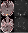
Post-contrast MR fused with MR venography (AC) and MR venography (BD). (A,B): thrombosis of the right transverse sinus (1), venography detects residual flow along its anterior wall (2). The left transverse sinus (TS) is hypoplastic. A prominent superior ophthalmic vein is shown on the right side (SOV). The left optic nerve (ON) is displaced by the intraorbital meningocele (M). (C,D): Fluid collection at the right mastoid with thrombosis of the sigmoid sinus (SS) and internal jugular vein (IJV). The right inferior petrosal sinus is patent (IPS). ICA: internal carotid artery.
Figure 3
Figure 3
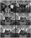
(AF): The coronal T2 sequence shows the intraorbital meningocele (M) displacing the optic nerve (ON) upward and medially (A). A prominent subarachnoid space at the level of both optic sheaths is present. The meningocele enters the orbit via the superior orbital fissure (SOF) (B). At the anterior aspect of the cavernous sinus (C), the meningocele runs below the anterior clinoid (AC) and above the maxillary groove of the maxillary nerve (V2); it displaces the lateral wall of the cavernous sinus (DF), causing a more convex shape (CS). At the posterior aspect of the cavernous sinus, the fluid signal is present both medially to the left internal carotid artery (white arrow) and laterally. The left oculomotor nerve runs in a lower position (IIIn) than the right one.
Figure 4
Figure 4
The sagittal T1 sequence shows the sella turcica filled by CSF with a distinct concavity of the hypophysis.
Figure 5
Figure 5
In the two T2 axial planes, the CSF signal is detected within the cavernous sinus, both medially and laterally to the internal carotid artery (white arrows on (A)). (B): The cisternal segment of the trigeminal nerve is eccentric with respect to the Meckel cave. A separation of the Meckel cave and cavernous sinus is not detectable, and the path of the CSF signal runs toward the superior orbital fissure ending in the intraorbital meningocele (black arrows).
Figure 6
Figure 6
Arteriography with venous phase. (A): Selective injection in the right ICA, a stent has been placed inside the right transverse sinus. (B): Patency of the stent is demonstrated. Residual stenosis of the right transverse sinus at the origin (white arrows). Hypoplastic left transverse sinus with faint contrast agent filling of the left jugular vein (black arrows). (C): The oblique view demonstrates a residual thrombus at the origin of the right jugular vein.
Figure 7
Figure 7
(A): CT obtained a few days after placement of the right transverse sinus stent shows residual thrombus (black arrow) and a reduction in the size of the intraorbital meningocele (white arrows). (B): At follow-up CT acquired after one month, complete patency of sigmoid sinus is achieved (black arrow). A further reduction in the size of the meningocele is shown (white arrows).
Figure 8
Figure 8
The clinical course of the patient.
Figure 9
Figure 9
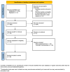
PRISMA flowchart illustrating the article selection process.
Figure 10
Figure 10
Bar plots of general characteristics of the systematic review: (A) ICH assessment; (B) herniation point; (C) surgery or interventional procedures performed; (D) outcome of ocular function.

References

    1. Flint P.W., Haughey B.H., Lund V.J., Robbins K.T., Thomas J.R., Lesperance M.M., Francis H.W. Cummings Otolaryngology–International Edition: Head and Neck Surgery. [(accessed on 19 January 2024)];2020 Volume 7:1323–1344. Available online: https://www.worldcat.org/title/1239324944.
    1. Suwanwela C., Suwanwela N. A morphological classification of sincipital encephalomeningoceles. J. Neurosurg. 1972;36:201–211. doi: 10.3171/jns.1972.36.2.0201. - DOI - PubMed
    1. Van Rumund A., Verrips A., Verhagen W.I.M. Pulsatile Proptosis due to Intraorbital Meningocele. Front. Neurol. 2017;8:290. doi: 10.3389/fneur.2017.00290. - DOI - PMC - PubMed
    1. Sadashivam S. Isolated Orbital Roof Fracture: Can It Be Catastrophic? Asian J. Neurosurg. 2018;13:935. doi: 10.4103/ajns.AJNS_101_18. - DOI - PMC - PubMed
    1. Gelabert-González M., García-Allut A., Villa J.F., Martínez-Rumbo R. Orbital meningocystocele. Neurocirugia. 2003;14:145–147; discussion 148. doi: 10.1016/S1130-1473(03)70552-1. - DOI - PubMed

LinkOut - more resources