. 2024 May 17;17(10):2413.
doi: 10.3390/ma17102413.

A Multidisciplinary Evaluation of Three-Dimensional Polycaprolactone Bioactive Glass Scaffolds for Bone Tissue Engineering Purposes

Affiliations

A Multidisciplinary Evaluation of Three-Dimensional Polycaprolactone Bioactive Glass Scaffolds for Bone Tissue Engineering Purposes

Gregorio Marchiori et al. Materials (Basel). .

Abstract

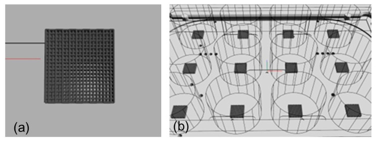
In the development of bone graft substitutes, a fundamental step is the use of scaffolds with adequate composition and architecture capable of providing support in regenerative processes both on the tissue scale, where adequate resistance to mechanical stress is required, as well as at the cellular level where compliant chemical-physical and mechanical properties can promote cellular activity. In this study, based on a previous optimization study of this group, the potential of a three-dimensional construct based on polycaprolactone (PCL) and a novel biocompatible Mg- and Sr-containing glass named BGMS10 was explored. Fourier-transform infrared spectroscopy and scanning electron microscopy showed the inclusion of BGMS10 in the scaffold structure. Mesenchymal stem cells cultured on both PCL and PCL-BGMS10 showed similar tendencies in terms of osteogenic differentiation; however, no significant differences were found between the two scaffold types. This circumstance can be explained via X-ray microtomography and atomic force microscopy analyses, which correlated the spatial distribution of the BGMS10 within the bulk with the elastic properties and topography at the cell scale. In conclusion, our study highlights the importance of multidisciplinary approaches to understand the relationship between design parameters, material properties, and cellular response in polymer composites, which is crucial for the development and design of scaffolds for bone regeneration.

Keywords: PCL; bioactive glasses; bone; composite scaffolds; human bone-marrow-derived mesenchymal stem cells; magnesium; therapeutic ions; tissue engineering.


Conflict of interest statement

Author Mauro Petretta was employed by the company REGENHU SA. The remaining authors declare that the research was conducted in the absence of any commercial or financial relationships that could be construed as a potential conflict of interest.

Figures

Figure 1
Figure 1
(a) design of the PCL-BGMS10 scaffolds. The red line is the axis origin while black line indicates landmarks on the building plate (b) Automated printing in 12-well plates. Blue-red lines are the axis origin, while black dots indicate reference points on the building plate.
Figure 2
Figure 2
Macroscopic pictures of the fabricated scaffolds. (a) 12-well plate. (b) Single scaffold close-up.
Figure 3
Figure 3
(a) Optical image of the microfibers deposited onto a glass slide and used for FTIR and AFM analyses. (b) Optical image showing the orientation of the microfiber with respect to the scan directions of the AFM measurement.
Figure 4
Figure 4
FTIR spectra of (ac) precursor material: PCL-BGMS10 (black line) and PCL (charcoal line) pellets and BGMS10 powder (light gray line) and 4 (df) printed fibers of PCL-BGMS10 (black line) and PCL (charcoal line). In (a) for PCL pellets are reported the symmetric (υs) and asymmetric (υas) vibrations discussed in the manuscript. The dashed boxers zoom (b,e) the methylene asymmetric (υasCH2) and symmetric (υsCH2) stretchings between 3100 and 2800 cm−1 and (c,f) the bands related to the crystalline phase (υcr) and the asymmetric (υasC-O-C) and symmetric (υsC-O-C) ester stretching between 1500 and 400 cm−1.
Figure 5
Figure 5
(a,b) Representative SEM micrographs of the PCL and (ce) PCL/BGMS10 scaffolds. (e,f) Results of the X-ray microanalysis performed at the point reported in (e).
Figure 6
Figure 6
Tukey box plot showed gene expression at 7, 14, and 21 days of the principal osteogenic markers. Different patterns were used for different scaffolds: PCLBGMS10 blue, PCL orange. * p < 0.05.
Figure 7
Figure 7
Transversal sections of (a) PCL scaffold and (b) PCL-BGMS10scaffold, mapping pore size (2D parameter “Size (ECDa)”) with colors and percentage on surface area (S,%) histograms.
Figure 8
Figure 8
Fiber thickness mapping of a PCL (a) and PCL-BGMS10 (b) scaffold, 3D rendered by SkyScan CTVox software (version 3.3.1).
Figure 9
Figure 9
BGMS10 particles’ size (a) distribution of the two PCL-BGMS10 samples (#1 and #2) and (b) mapping of a sample, 3D rendered by SkyScan CTVox software. Eventual particles below 5 µm cannot be seen because of MicroCT spatial resolution.
Figure 10
Figure 10
% of BGMS10 content expressed as (mean ± SD) of the two samples, in concentric ROIs mapped on a PCL-BGMS10 scaffold, 3D rendered by SkyScan CTVox software.
Figure 11
Figure 11
MicroCT image dataset gray-scale histograms (0–255 gray levels) showing (a) greater areas with high gray levels (180–255, red arrow) for a PCL scaffold harvested for 21 days (PCL_21dCells) respect to the baseline (PCL_no cells) and (b) greater areas with low gray levels (12–60, red arrow) for PCL-BGMS10 scaffolds harvested for 7/21 days (PCL-BGMS10_7dCells/PCL-BGMS10_21dCells) respect to the baseline (PCL-BGMS10 no cells).
Figure 12
Figure 12
Sagittal cut of MicroCT image reconstruction (left) and corresponding gray-scale histogram (right) of (a) PCL scaffold harvested for 21 days (PCL_21dCells) and (b) PCL-BGMS10 scaffold harvested for 21 days (PCL-BGMS10_21dCells), showing possible cellular deposit (in pink or red in sagittal cut images, with relative gray levels highlighted in histograms) on the scaffold (in green in sagittal cut images, gray levels not highlighted in histograms) surfaces.
Figure 13
Figure 13
Topographic 10 µm × 4 µm images of (a) PCL and (b) PCL-BGMS10.
Figure 14
Figure 14
(a) Schematic of the AFM-based nanoindentation and (b) Statistical distribution of Young’s moduli extracted from 1440 nanoindentation points taken on the topographic regions of Figure 13a (blue bars) and Figure 13b (red bars) and the corresponding Gaussian fittings (software OriginPro version 7.5).

References

    1. Florencio-Silva R., Sasso G.R.D.S., Sasso-Cerri E., Simões M.J., Cerri P.S. Biology of Bone Tissue: Structure, Function, and Factors That Influence Bone Cells. Biomed. Res. Int. 2015;2015:421746. doi: 10.1155/2015/421746. - DOI - PMC - PubMed
    1. Dwivedi R., Kumar S., Pandey R., Mahajan A., Nandana D., Katti D.S., Mehrotra D. Polycaprolactone as Biomaterial for Bone Scaffolds: Review of Literature. J. Oral Biol. Craniofacial Res. 2020;10:381–388. doi: 10.1016/j.jobcr.2019.10.003. - DOI - PMC - PubMed
    1. Judex S., Carlson K.J. Is Bone’s Response to Mechanical Signals Dominated by Gravitational Loading? Med. Sci. Sports Exerc. 2009;41:2037–2043. doi: 10.1249/MSS.0b013e3181a8c6e5. - DOI - PubMed
    1. Lee S.S., Du X., Kim I., Ferguson S.J. Scaffolds for Bone-Tissue Engineering. Matter. 2022;5:2722–2759. doi: 10.1016/j.matt.2022.06.003. - DOI
    1. Gharibshahian M., Salehi M., Beheshtizadeh N., Kamalabadi-Farahani M., Atashi A., Nourbakhsh M.S., Alizadeh M. Recent Advances on 3D-Printed PCL-Based Composite Scaffolds for Bone Tissue Engineering. Front. Bioeng. Biotechnol. 2023;11:1168504. doi: 10.3389/fbioe.2023.1168504. - DOI - PMC - PubMed