. 2024 Apr 29;16(5):708.
doi: 10.3390/v16050708.

Development of a Cell Culture Model for Inducible SARS-CoV-2 Replication

Affiliations

Development of a Cell Culture Model for Inducible SARS-CoV-2 Replication

Xiaoyan Wang et al. Viruses. .

Abstract

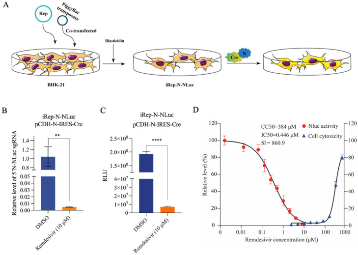
Severe acute respiratory syndrome coronavirus 2 (SARS-CoV-2) induces direct cytopathic effects, complicating the establishment of low-cytotoxicity cell culture models for studying its replication. We initially developed a DNA vector-based replicon system utilizing the CMV promoter to generate a recombinant viral genome bearing reporter genes. However, this system frequently resulted in drug resistance and cytotoxicity, impeding model establishment. Herein, we present a novel cell culture model with SARS-CoV-2 replication induced by Cre/LoxP-mediated DNA recombination. An engineered SARS-CoV-2 transcription unit was subcloned into a bacterial artificial chromosome (BAC) vector. To enhance biosafety, the viral spike protein gene was deleted, and the nucleocapsid gene was replaced with a reporter gene. An exogenous sequence was inserted within NSP1 as a modulatory cassette that is removable after Cre/LoxP-mediated DNA recombination and subsequent RNA splicing. Using the PiggyBac transposon strategy, the transcription unit was integrated into host cell chromatin, yielding a stable cell line capable of inducing recombinant SARS-CoV-2 RNA replication. The model exhibited sensitivity to the potential antivirals forsythoside A and verteporfin. An innovative inducible SARS-CoV-2 replicon cell model was introduced to further explore the replication and pathogenesis of the virus and facilitate screening and assessment of anti-SARS-CoV-2 therapeutics.

Keywords: inducible model; replicon; severe acute respiratory syndrome coronavirus 2.


Conflict of interest statement

The authors declare no conflicts of interest.

Figures

Figure 1
Figure 1
Design of the Rep-S-EGFP/NLuc replicon. (A) Design of the SARS-CoV-2 viral genome structure and replicon model. We selectively excised a portion of the S gene coding sequence while preserving its transcriptional regulatory sequence (TRS) and inserted the “self-cleaving peptide” T2A-linked EGFP or IgK secretion signal peptide-tagged NLuc sequences, allowing NLuc or EGFP to be expressed freely. The CMV promoter and hepatitis D virus anti-ribozyme sequence (HDV RZ) were introduced to the 5′ and 3′ ends of the SARS-CoV-2 genome, respectively, to construct the transcription unit. (B) Flowchart of the SARS-CoV-2 recombinant replicon assembly strategy. Utilizing a bacterial artificial chromosome (pBAC) as the vector, the pBAC-CMV-5′UTR-EGFP/NLuc-N-3′UTR precursor clone was generated using fusion PCR. This precursor clone was subsequently digested with restriction enzymes and ligated with ORF1ab segments (F1/F2/F3/F4) harboring homologous arms, culminating in the assembly of the replicon clone plasmid Rep-S-EGFP/NLuc using Gibson assembly.
Figure 2
Figure 2
Rep-S-EGFP replication in transfected cells. (A) Changes in nucleocapsid (N) protein levels in Rep-S-EGFP-transfected cells and the effect of remdesivir on its expression. The Rep-S-EGFP replicon plasmid was transfected into VeroE6 and Huh7.5 cells. After 6 h, 10 μM remdesivir was added. Cell lysates were collected at 24, 48, 60, and 72 h post-transfection for western blotting analysis. (B) At 48 h post-transfection, VeroE6 cells as depicted in (A) were imaged under a fluorescence microscope. Representative images from three independent experiments are shown. (C) Schematic representation of qRT-PCR primers N-sg-F and N-sg-R targeting N gene sgRNAs. The replication of SARS-CoV-2 generates numerous subgenomic RNAs, as indicated in the diagram. (D) Rep-S-EGFP replicon-transfected BHK-21 cells were treated with remdesivir at indicated concentrations for 36 h. N gene sgRNA was quantified using qRT-RCR with specific primers. Each group included 3 biological replicates. * indicates p < 0.05, ** indicates p < 0.01, *** indicates p < 0.001. (E) The Rep-S-EGFP replicon plasmid-transfected BHK-21 cells were treated with different concentrations of remdesivir for 36 h. N sgRNA was determined as depicted in (D). Cytotoxicity was evaluated using the CCK-8 assay. Cells mock-treated with DMSO were used as control. Selectivity index (value of CC50/IC50, SI). (F) Differential expression of progeny N sgRNAs between the replicons Rep-S-EGFP and Rep-S-EGFP-RdRpmut. Data were normalized to the Rep-S-EGFP-RdRpmut control. *** indicates p < 0.001. (G) Comparison of N protein levels in Rep-S-EGFP- or Rep-S-EGFP-RdRpmut-transfected BHK-21 cells. Treated with remdesivir for 36 h after transfection for 6 h. Cell samples were collected and analyzed using western blotting. (H) Rep-S-EGFP replicon-transfected BHK-21 cells were treated with nirmatrelvir at indicated concentrations for 36 h. The inhibitory effect and cytotoxicity of nirmatrelvir on Rep-S-EGFP were investigated as depicted in (E). (I) The N protein expression in nirmatrelvir-treated cells, as described in (H), was determined using western blotting.
Figure 3
Figure 3
Impact of remdesivir on reporter genes in Rep-S-NLuc and optimized Rep-N-NLuc replicons. (A) Inhibitory effect of remdesivir on Rep-S-NLuc NLuc activity. The Rep-S-NLuc plasmid was co-transfected with a Renilla luciferase-expressing plasmid into VeroE6, Huh7.5, and BHK-21 cells. After a 6-hour transfection, 10 μM remdesivir was added. Cell lysates were collected at 36 h post-transfection for NLuc activity analysis. Each treatment group included three biological replicates. Data were normalized to that of Renilla and analyzed for differential expression, with the DMSO group serving as the control. Results are presented as the mean ± SEM. *** indicates p < 0.001, **** indicates p < 0.0001. (B) Optimization of the reporter gene position for the Rep-N-NLuc replicon plasmid. The N gene fragment was deleted while retaining its upstream transcription regulatory sequence (TRS). The non-secreted NLuc gene was inserted into the deleted location to rescue viral replication using N gene complementation. (C) Remdesivir significantly inhibited the NLuc activity (relative luminescence units, RLU) of the Rep-N-NLuc replicon. The Rep-N-NLuc replicon plasmid, pcDNA3.1-N, and Renilla luciferase-expressing plasmid were co-transfected into BHK-21 cells. The deep blue bar indicates the DMSO-treated MOCK group, while the orange bar represents the group treated with 10 μM remdesivir.Each treatment group included three biological replicates. *** indicates p < 0.001. (D) Remdesivir significantly inhibited the generation of N-NLuc sgRNAs in Rep-N-NLuc. qRT-PCR detection was performed using the samples from (C). N-NLuc sgRNA was detected using special primers. ** indicates p < 0.01.
Figure 4
Figure 4
An inducible SARS-CoV-2 replicon system co-transfected with N-protein rescued viral replication upon Cre/loxP-mediated site-specific recombination. (A) Structure of the recombinant SARS-CoV-2 replicon (iRep-N-NLuc). A transcription stop cassette integrated between nt 66 and 67 (i.e., CAG66^G67; ^ indicates the potential exon/exon boundary) of the NSP1 gene. The inserted sequence consists of the indicated elements in order. PiggyBac 5′ TR and chicken β-globin insulators are inserted at the 5′ end of the CMV promoter and at the 3′ end of the HDV RZ region, respectively, flanking the transcription unit. “L” stands for NLuc. (B) A schema of the intracellular process of inducible iRep-N-NLuc replication in the cell model. In the absence of Cre recombinase, the CMV promoter only drives the expression of the blasticidin resistance gene, preventing SARS-CoV-2 replication, facilitating the selection of stably integrated cell lines. Following Cre recombinase induction in replicon cell lines, site-specific recombination removes the transcription stop cassette at the DNA level. The CMV promoter then initiates transcription of the viral genome. RNA splicing mechanisms within the nucleus eliminate the remaining single LoxP site. The cell nucleus is depicted in blue, while the cytoplasm is shown in light blue. (C) BHK-21 cells were co-transfected with the iRep-N-NLuc replicon and indicated plasmids plus a Renilla luciferase normalization plasmid. The fold change in NLuc activity was normalized to that of the Renilla luciferase activity. *** indicates p < 0.001. (D) BHK-21 cells were co-transfected with iRep-N-NLuc replicon, pCDNA3.1-N, and pCDH-Cre. At 6 h after transfection, cells were treated with 10 μM remdesivir for an additional 24 h and subjected to analysis of NLuc activity. Cells mock-treated with DMSO were used as a control. ** indicates p < 0.01.
Figure 5
Figure 5
Generation of an inducible stable cell line producing a recombinant SARS-CoV-2 replicon. (A) Schematic illustration of the procedure to generate a cell-based SARS-CoV-2 replicon model. (B,C) The iRep-N-NLuc cell line was transfected with a bicistronic IRES vector (pCDH-N-IRES-Cre). Cells were either treated with 10 μM remdesivir or mock treated with DMSO. The recombinant N-NLuc sgRNAs (B) and NLuc activity (C) were determined at 24 h post-transfection. Error bars indicate the mean ± SEM from three independent experiments. ** indicates p < 0.01, **** indicates p < 0.0001. (D) pCDH-N-IRES-Cre-transfected iRep-N-NLuc cells were treated with different concentrations of remdesivir for 24 h. NLuc activity was determined. Cytotoxicity was evaluated using the CCK-8 assay. Data were expressed as the percentage of the mock-treated controls. Error bars indicate the mean ± SEM from three independent experiments.
Figure 6
Figure 6
Antiviral drug screening and validation based on the inducible SARS-CoV-2 replicon cell model. (A) Schematic illustration of the procedure for antiviral screening based on the iRep-N-NLuc cells model. (B) Evaluation of the antiviral activity of candidate compounds in the iRep-N-NLuc-cells model. Cells were transfected with pCDH-N-IRES-Cre and treated with candidate compounds at indicated doses. The NLuc activity was determined at 24 h post-transfection and were expressed as a percentage of the values measured in DMSO-treated cells. Error bars indicate the mean ± SEM from three independent experiments. ** indicates p < 0.01, *** indicates p < 0.001, **** indicates p < 0.0001. (C) iRep-N-NLuc cells were transfected with pCDH-N-IRES-Cre, followed by the treatment with forsythiaside A at indicated doses for 24 h. NLuc activity was determined as depicted in (B). Cell viability was measured using the CCK-8 assay. (D) BHK-21 cells were transfected with Rep-S-EGFP and treated with forsythiaside A at indicated doses. After 48 h, N protein expression was determined using western blotting. (E) Rep-N-NLuc-transfected cells were treated with varying concentrations of verteporfin for 24 h. NLuc activity was measured and expressed as a percentage of the values measured in mock-treated cells. Cell cytotoxicity and CC50 were assessed using the CCK-8 assay. (F) Rep-S-GFP-transfected cells were treated with verteporfin for 36 h. N sgRNA was determined using qRT-PCR. Data are presented as the mean ± SEM from three independent experiments. ** indicates p < 0.01. (G) iRep-N-NLuc cells were transfected with pCDH-N-IRES-Cre for 6 h and subsequently treated with varying concentrations of nirmatrelvir. NLuc activity was determined as depicted in (B). The cytotoxicity assay is the same as the CCK8 assay in Figure 2G.

Similar articles

Cited by

References

    1. Chow E.J., Uyeki T.M., Chu H.Y. The effects of the COVID-19 pandemic on community respiratory virus activity. Nat. Rev. Microbiol. 2023;21:195–210. doi: 10.1038/s41579-022-00807-9. - DOI - PMC - PubMed
    1. Li G., Hilgenfeld R., Whitley R., De Clercq E. Therapeutic strategies for COVID-19: Progress and lessons learned. Nat. Rev. Drug Discov. 2023;22:449–475. doi: 10.1038/s41573-023-00672-y. - DOI - PMC - PubMed
    1. Gordon C.J., Tchesnokov E.P., Schinazi R.F., Götte M. Molnupiravir promotes SARS-CoV-2 mutagenesis via the RNA template. J. Biol. Chem. 2021;297:100770. doi: 10.1016/j.jbc.2021.100770. - DOI - PMC - PubMed
    1. Fernandes R.S., Freire M., Bueno R.V., Godoy A.S., Gil L., Oliva G. Reporter Replicons for Antiviral Drug Discovery against Positive Single-Stranded RNA Viruses. Viruses. 2020;12:598. doi: 10.3390/v12060598. - DOI - PMC - PubMed
    1. Zhang Y., Song W., Chen S., Yuan Z., Yi Z. A bacterial artificial chromosome (BAC)-vectored noninfectious replicon of SARS-CoV-2. Antivir. Res. 2021;185:104974. doi: 10.1016/j.antiviral.2020.104974. - DOI - PMC - PubMed

LinkOut - more resources