. 2024 May 11;16(5):761.
doi: 10.3390/v16050761.

User-Friendly Replication-Competent MAdV-1 Vector System with a Cloning Capacity of 3.3 Kilobases

Affiliations

User-Friendly Replication-Competent MAdV-1 Vector System with a Cloning Capacity of 3.3 Kilobases

Zhichao Zhang et al. Viruses. .

Abstract

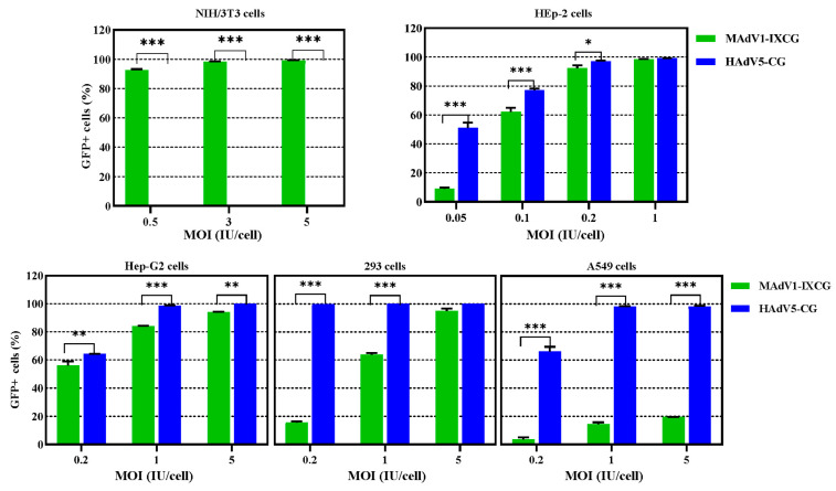
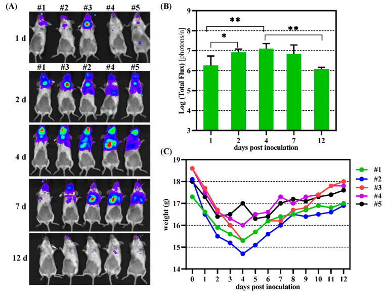
Mouse adenoviruses (MAdV) play important roles in studying host-adenovirus interaction. However, easy-to-use reverse genetics systems are still lacking for MAdV. An infectious plasmid pKRMAV1 was constructed by ligating genomic DNA of wild-type MAdV-1 with a PCR product containing a plasmid backbone through Gibson assembly. A fragment was excised from pKRMAV1 by restriction digestion and used to generate intermediate plasmid pKMAV1-ER, which contained E3, fiber, E4, and E1 regions of MAdV-1. CMV promoter-controlled GFP expression cassette was inserted downstream of the pIX gene in pKMAV1-ER and then transferred to pKRMAV1 to generate adenoviral plasmid pKMAV1-IXCG. Replacement of transgene could be conveniently carried out between dual BstZ17I sites in pKMAV1-IXCG by restriction-assembly, and a series of adenoviral plasmids were generated. Recombinant viruses were rescued after transfecting linearized adenoviral plasmids to mouse NIH/3T3 cells. MAdV-1 viruses carrying GFP or firefly luciferase genes were characterized in gene transduction, plaque-forming, and replication in vitro or in vivo by observing the expression of reporter genes. The results indicated that replication-competent vectors presented relevant properties of wild-type MAdV-1 very well. By constructing viruses bearing exogenous fragments with increasing size, it was found that MAdV-1 could tolerate an insertion up to 3.3 kb. Collectively, a replication-competent MAdV-1 vector system was established, which simplified procedures for the change of transgene or modification of E1, fiber, E3, or E4 genes.

Keywords: Gibson assembly; infectious plasmid; mouse adenovirus 1; packaging capacity; replication competent; transgene; vector.


Conflict of interest statement

The authors declare no conflicts of interest. The funders had no role in the design of the study, in the collection, analyses, or interpretation of data, in the writing of the manuscript, or in the decision to publish the results.

Figures

Figure 1
Figure 1
Schematic diagram of the construction of replication-competent MAdV-1 vector. An intermediate plasmid-based strategy was used to modify the genome of MAdV-1. pKRMAV1 was an infectious plasmid that contained the whole MAdV-1 genome. Intermediate plasmid pKMAV1-ER was generated by fusing the EcoRI/RsrII-digesting product of pKRMAV1 and other fragments of PCR products with Gibson assembly ①. pKMAV1-ER was a small plasmid with more unique restriction sites that could be used for site-directed mutation. CMV promoter-controlled GFP expression cassette was inserted downstream of the pIX gene to generate pKMAV1-ERCG ②. The modified intermediate plasmid of pKMAV1-ERCG was brought back to pKRMAV1 to generate the final adenoviral plasmid of pKMAV1-IXCG by restriction-assembly ③④⑤. The details can be found in the Materials and Methods section.
Figure 2
Figure 2
Rescue and identification of MAdV1-IXCG recombinant virus. (A) Rescue of MAdV1-IXCG virus. PmeI-linearized pKMAdV1-IXCG was used to transfect NIH/3T3 cells. The expression of GFP was observed under a fluorescence microscope. The occurrence and growth of GFP foci implied that the MAdV1-IXCG virus was successfully rescued. (B) Identification of MAdV1-IXCG by restriction analysis of its genomic DNA. Virus genomic DNA (G) was digested with the indicated restriction enzymes and resolved on 0.7% agarose gel by electrophoresis, and purified DNA of adenoviral plasmid pKMAdV1-IXCG (P) served as a control. The predicted molecular weights (bp) of digested fragments of MAdV1-IXCG genome were 1102, 2354, 3381, 4673, 8056, 12,382 for AseI; 1112, 1284, 1875, 3692, 10,735, 12,690 for KpnI; 1297, 1908, 2106, 2188, 5643, 6997, 11,866 for NdeI; and 3887, 4606, 5640, 7268, 10,844 for SacII. The predicted molecular weights (bp) of digested fragments of pKMAdV1-IXCG plasmid were 1102, 2354, 3381, 5818, 9382, 12,382 for AseI; 1112, 1875, 3692, 10,735, 16,445 for KpnI; 1297, 1908, 5643, 6765, 6997, 11,866 for NdeI; and 4606, 5640, 7268, 17,202 for SacII. Molecular weights of fragments less than 1000 bp were not given.
Figure 3
Figure 3
Transduction ability of replication-competent MAdV-1 vector carrying GFP reporter gene. Mouse NIH/3T3 and human adherent cell lines were infected with MAdV1-IXCG or control HAdV5-CG for 4 h at various MOIs; the cells were harvested 2 days post-infection, and the expression of GFP was assayed by flow cytometry. All the experiments were performed in duplicate, and the data shown are from one representative experiment out of the two performed. * p < 0.05, ** p < 0.01 and *** p < 0.001.
Figure 4
Figure 4
Growth of MAdV1-IXCG in packaging NIH/3T3 cells. (A) Plaque forming experiments. NIH3T3 cells in 6-well plates were infected with MAdV1-IXCG or the parental MAdV-1 at low doses for 2 h and then cultured in a semi-solid culture medium. The cells were fixed in paraformaldehyde, and the plaques were visualized by crystal violet staining 6 days post-infection. (B) The areas of total foci in one well were measured for each virus using the Fiji image processing package. The violin plots of the focus size data are shown. The size medians were compared by using the Kruskal–Wallis non-parameter test. (C) One-step growth curve. NIH/3T3 cells in 12-well plates were infected with MAdV1-IXCG or MAdV-1 at an MOI of 2 IU/cell for 4 h. Culture media and infected cells were harvested at indicated time points and used for titration. The yields of progeny viruses associated with cells or released to the culture medium were calculated, respectively.
Figure 5
Figure 5
Replication of MAdV1-IXCG genome or infectious virus in human HEp-2 cells. (A) Mouse NIH3T3 or human HEp-2 cells in 12-well plates were infected in duplicate with MAdV1-IXCG at an MOI of 0.5 IU/cell for 4 h. Virus diluents were removed, and cells were washed twice before fresh maintenance media were added. Cells were collected at indicated time points, and total DNA was extracted. TaqMan probe-based real-time PCR was performed in triplicate to determine the copy number of the viral genome. The copy numbers of the viral genome in the well were calculated and shown. (B) NIH/3T3 or HEp-2 cells in 12-well plates were similarly infected with MAdV1-IXCG at MOIs of 0.5 or 5 IU/cell. At indicated time points, the cells, together with the culture medium, were harvested and used for titration. LOD—limit of detection; ud—undetected.
Figure 6
Figure 6
Biodistribution of MAdV1-IXCG in mice after intranasal administration. (A) The mice were intranasally infected with MAdV1-IXCLG at a dose of 1.2 × 10⁴ IU per mouse. In vivo bioluminescence imaging was performed to detect the activity of firefly luciferase in mice on sequential days post-virus inoculation. (B) Total photon flux measured after drawing the regions of interest (ROIs) in bioluminescence images. (C) Body weights of mice monitored on consecutive days post-infection. * p < 0.05, ** p < 0.01.
Figure 7
Figure 7
Schematic diagram of the transgene expression cassettes with gradually increasing size of stuffer DNA in recombinant MAdV-1 viruses. The details of the construction of these viruses were described in the Materials and Methods section. CMVp—CMV promoter; pA—polyA signal. IXCG, CG1K, CG2K, CG3K, CG4K, and CG5K were short names for MAdV1-IXCG, MAdV1-IXCG1K, MAdV1-IXCG2K, MAdV1-IXCG3K, MAdV1-IXCG4K, and MAdV1-IXCG5K.
Figure 8
Figure 8
Growth of recombinant MAdV-1 viruses with increasing sizes of carried exogenous DNA. (A) Virus rescue in packaging cells. PmeI-linearized adenoviral plasmids were used to transfect NIH/3T3 cells. GFP-positive cells and occurrence of GFP foci were monitored by observing under a fluorescence microscope (MAdV1-IXCG5K failed in rescue). (B) Violin plots of the plaque size data. Rescued recombinant viruses and MAdV1-IXCG were subjected to plaque-forming experiments. The cells were fixed and stained 6 days post-infection. The areas of all plaques formed in one well were measured for each virus. The data were analyzed using the nonparametric Kruskal–Wallis test, and mean ranks were compared between two neighboring viruses. ** p < 0.01; *** p < 0.001. (C) Growth of recombinant viruses in fully infected packaging cells. NIH/3T3 cells in a 12-well plate were infectious with recombinant viruses at an MOI of 1 IU/cell for 4 h. The culture media and cells were harvested 48 or 96 h post-infection and used for titration. Virus yields were calculated. IXCG, CG1K, CG2K, CG3K, and CG4K were short names for MAdV1-IXCG, MAdV1-IXCG1K, MAdV1-IXCG2K, MAdV1-IXCG3K, and MAdV1-IXCG4K.

Similar articles

Cited by

References

    1. Benkő M., Aoki K., Arnberg N., Davison A.J., Echavarría M., Hess M., Jones M.S., Kaján G.L., Kajon A.E., Mittal S.K., et al. ICTV Virus Taxonomy Profile: Adenoviridae 2022. J. Gen. Virol. 2022;103:001721. doi: 10.1099/jgv.0.001721. - DOI - PMC - PubMed
    1. Crystal R.G. Adenovirus: The First Effective In Vivo Gene Delivery Vector. Hum. Gene Ther. 2014;25:3–11. doi: 10.1089/hum.2013.2527. - DOI - PMC - PubMed
    1. Sakurai F., Tachibana M., Mizuguchi H. Adenovirus Vector-Based Vaccine for Infectious Diseases. Drug Metab. Pharmacokinet. 2022;42:100432. doi: 10.1016/j.dmpk.2021.100432. - DOI - PMC - PubMed
    1. Trivedi P.D., Byrne B.J., Corti M. Evolving Horizons: Adenovirus Vectors’ Timeless Influence on Cancer, Gene Therapy and Vaccines. Viruses. 2023;15:2378. doi: 10.3390/v15122378. - DOI - PMC - PubMed
    1. Lundstrom K. Viral Vectors in Gene Therapy: Where Do We Stand in 2023? Viruses. 2023;15:698. doi: 10.3390/v15030698. - DOI - PMC - PubMed

LinkOut - more resources