Review
. 2024 Apr 27;12(5):467.
doi: 10.3390/vaccines12050467.

The D Gene in CDR H3 Determines a Public Class of Human Antibodies to SARS-CoV-2

Affiliations
Review

The D Gene in CDR H3 Determines a Public Class of Human Antibodies to SARS-CoV-2

Meng Yuan et al. Vaccines (Basel). .

Abstract

Public antibody responses have been found against many infectious agents. Structural convergence of public antibodies is usually determined by immunoglobulin V genes. Recently, a human antibody public class against SARS-CoV-2 was reported, where the D gene (IGHD3-22) encodes a common YYDxxG motif in heavy-chain complementarity-determining region 3 (CDR H3), which determines specificity for the receptor-binding domain (RBD). In this review, we discuss the isolation, structural characterization, and genetic analyses of this class of antibodies, which have been isolated from various cohorts of COVID-19 convalescents and vaccinees. All eleven YYDxxG antibodies with available structures target the SARS-CoV-2 RBD in a similar binding mode, where the CDR H3 dominates the interaction with antigen. The antibodies target a conserved site on the RBD that does not overlap with the receptor-binding site, but their particular angle of approach results in direct steric hindrance to receptor binding, which enables both neutralization potency and breadth. We also review the properties of CDR H3-dominant antibodies that target other human viruses. Overall, unlike most public antibodies, which are identified by their V gene usage, this newly discovered public class of YYDxxG antibodies is dominated by a D-gene-encoded motif and uncovers further opportunities for germline-targeting vaccine design.

Keywords: CDR H3; D gene; SARS-CoV-2; broadly neutralizing antibody; public antibody.


Conflict of interest statement

The authors declare no conflicts of interest.

Figures

Figure 1
Figure 1
ADI-62113 as a model for YYDxxG antibodies. (A) Conservation of the spike protein across SARS-CoV-2 variants (PDB 7SXZ). One protomer with RBD (receptor-binding domain) in its up conformation shows conservation, as calculated by ConSurf [14] and color-coded here. The ADI-62113 epitope is defined by its buried surface area (BSA > 0 Å2) on the RBD, as calculated by Proteins, Interfaces, Structures and Assemblies (PISA; www.ebi.ac.uk/pdbe/prot_int/pistart.html, accessed on 1 July 2023) and outlined. The other two protomers of the spike trimer are shown as a transparent grey surface. (B) The SARS-CoV-2 RBD is shown as a white surface, while the heavy and light chains of ADI-62113 are in orange and yellow, respectively (PDB 7T7B). The RBD–antibody structure is superimposed onto an RBD-ACE2 (angiotensin-converting enzyme 2) structure (PDB 6M0J) and illustrates that ADI-62113 would clash (red circle) with ACE2 (green). (C) Detailed interactions between the CDR H3 (heavy-chain complementarity-determining region 3) YYDxxG motif of ADI-62113 with SARS-CoV-2 RBD. Hydrogen bonds are indicated by dashed lines.
Figure 2
Figure 2
Structural comparison of YYDxxG antibodies. (A) All YYDxxG antibodies target SARS-CoV-2 RBD in a similar binding mode. The CDRs H3 (indicated by a red arrow in the first structure COVA1-16 as a representative) are responsible for the majority of the antigen interactions. The SARS-CoV-2 RBD is shown in whitish grey, while the heavy and light chains of the antibodies are in orange and yellow. For antibodies 2-36, VacW-209, and G32Q4, only the variable domain is available in the published structure. (B) Germline genes of the YYDxxG antibodies with available structures.
Figure 3
Figure 3
CR3022 and COVA1-16 target a similar epitope but with distinct angles of approach. (A) Epitopes and (B) structures of CR3022 (orange, PDB 6W41) and COVA1-16 (cyan, PDB 7JMW) in complex with the RBD (whitish grey) are depicted here. The epitopes are defined by the buried surface area (BSA > 0 Å2) on the RBD when bound to the antibody.
Figure 4
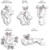
Figure 4
Representative antibodies with a dominant CDR H3 in complex with viral surface proteins. Variable domains of antibodies are represented by grey cartoon, and viral antigens by white surface. CDRs H3 are shown in red tubes. Only one CDR H3 is shown in red if multiple antibodies are bound in a viral protein.

References

    1. Fiolet T., Kherabi Y., MacDonald C.J., Ghosn J., Peiffer-Smadja N. Comparing COVID-19 vaccines for their characteristics, efficacy and effectiveness against SARS-CoV-2 and variants of concern: A narrative review. Clin. Microbiol. Infect. 2022;28:202–221. doi: 10.1016/j.cmi.2021.10.005. - DOI - PMC - PubMed
    1. Barnes C.O., West A.P., Jr., Huey-Tubman K.E., Hoffmann M.A.G., Sharaf N.G., Hoffman P.R., Koranda N., Gristick H.B., Gaebler C., Muecksch F., et al. Structures of human antibodies bound to SARS-CoV-2 spike reveal common epitopes and recurrent features of antibodies. Cell. 2020;182:828–842.e16. doi: 10.1016/j.cell.2020.06.025. - DOI - PMC - PubMed
    1. Yuan M., Liu H., Wu N.C., Lee C.D., Zhu X., Zhao F., Huang D., Yu W., Hua Y., Tien H., et al. Structural basis of a shared antibody response to SARS-CoV-2. Science. 2020;369:1119–1123. doi: 10.1126/science.abd2321. - DOI - PMC - PubMed
    1. Robbiani D.F., Gaebler C., Muecksch F., Lorenzi J.C.C., Wang Z., Cho A., Agudelo M., Barnes C.O., Gazumyan A., Finkin S., et al. Convergent antibody responses to SARS-CoV-2 in convalescent individuals. Nature. 2020;584:437–442. doi: 10.1038/s41586-020-2456-9. - DOI - PMC - PubMed
    1. Chi X., Yan R., Zhang J., Zhang G., Zhang Y., Hao M., Zhang Z., Fan P., Dong Y., Yang Y., et al. A neutralizing human antibody binds to the N-terminal domain of the Spike protein of SARS-CoV-2. Science. 2020;369:650–655. doi: 10.1126/science.abc6952. - DOI - PMC - PubMed

LinkOut - more resources