. 2024 May 7;12(5):505.
doi: 10.3390/vaccines12050505.

Antigenic Characterization of Circulating and Emerging SARS-CoV-2 Variants in the U.S. throughout the Delta to Omicron Waves

Affiliations

Antigenic Characterization of Circulating and Emerging SARS-CoV-2 Variants in the U.S. throughout the Delta to Omicron Waves

Han Di et al. Vaccines (Basel). .

Abstract

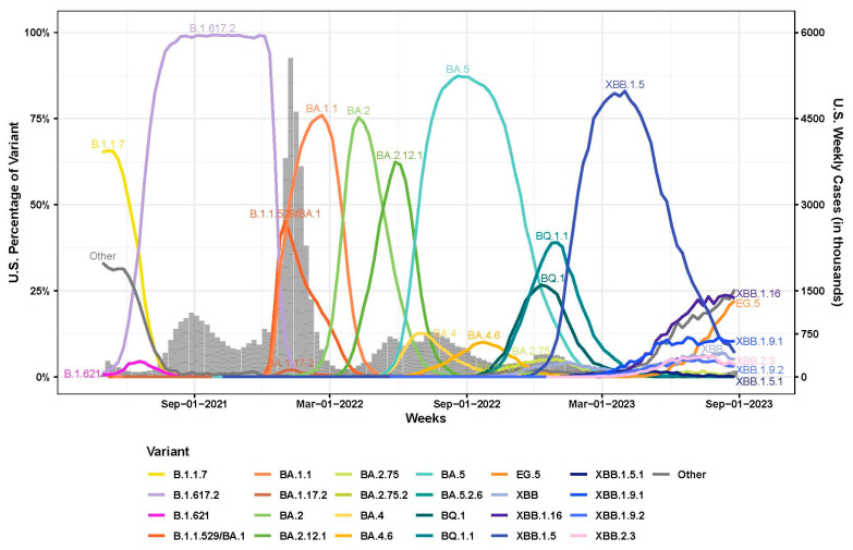
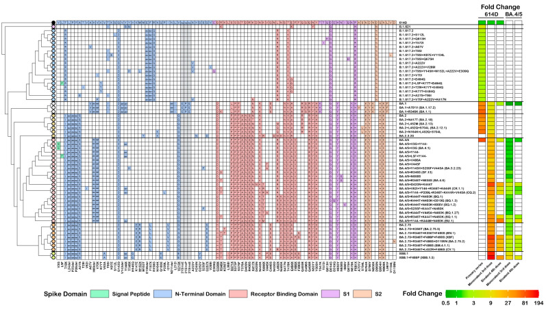
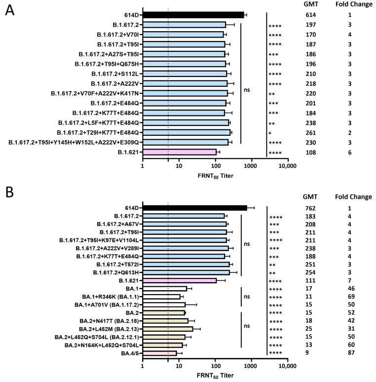
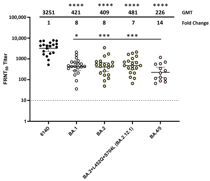
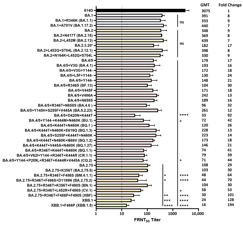
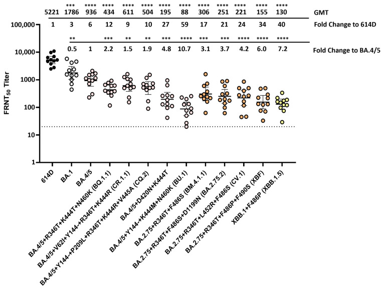
Severe acute respiratory syndrome coronavirus 2 (SARS-CoV-2) has evolved into numerous lineages with unique spike mutations and caused multiple epidemics domestically and globally. Although COVID-19 vaccines are available, new variants with the capacity for immune evasion continue to emerge. To understand and characterize the evolution of circulating SARS-CoV-2 variants in the U.S., the Centers for Disease Control and Prevention (CDC) initiated the National SARS-CoV-2 Strain Surveillance (NS3) program and has received thousands of SARS-CoV-2 clinical specimens from across the nation as part of a genotype to phenotype characterization process. Focus reduction neutralization with various antisera was used to antigenically characterize 143 SARS-CoV-2 Delta, Mu and Omicron subvariants from selected clinical specimens received between May 2021 and February 2023, representing a total of 59 unique spike protein sequences. BA.4/5 subvariants BU.1, BQ.1.1, CR.1.1, CQ.2 and BA.4/5 + D420N + K444T; BA.2.75 subvariants BM.4.1.1, BA.2.75.2, CV.1; and recombinant Omicron variants XBF, XBB.1, XBB.1.5 showed the greatest escape from neutralizing antibodies when analyzed against post third-dose original monovalent vaccinee sera. Post fourth-dose bivalent vaccinee sera provided better protection against those subvariants, but substantial reductions in neutralization titers were still observed, especially among BA.4/5 subvariants with both an N-terminal domain (NTD) deletion and receptor binding domain (RBD) substitutions K444M + N460K and recombinant Omicron variants. This analysis demonstrated a framework for long-term systematic genotype to antigenic characterization of circulating and emerging SARS-CoV-2 variants in the U.S., which is critical to assessing their potential impact on the effectiveness of current vaccines and antigen recommendations for future updates.

Keywords: COVID-19 vaccine; Delta variant; Omicron variant; SARS-CoV-2; antigenic characterization; neutralizing antibody.


Conflict of interest statement

H.H was supported by Synergy America, Inc. and S.H.P. was supported by Eagle Global Scientific, Inc. Synergy America, Inc. and Eagle Global Scientific, Inc. had no involvement in study design; collection, analysis, and interpretation of data; writing of the report and the decision to submit the report for publication.

Figures

Figure 1
Figure 1
Weekly proportions of SARS-CoV-2 variants in the U.S. with underlying U.S. case counts (1 May 2021 to 1 September 2023). Percentage of SARS-CoV-2 variants in the U.S. (line graph, 0% to 100%) were assembled from all available U.S. sequencing surveillance data. Variants represent an aggregation of many individual pangolin lineages aliased under a major parental lineage designation and were labeled with different colors. U.S. Weekly case counts were obtained from HHS Protect and are summarized in the grey bar graph (right-side, dual Y-axis).
Figure 2
Figure 2
Genetic and antigenic summary of the analyzed SARS-CoV-2 Delta, Mu and Omicron variants. Spike mutations relative to the 614D reference virus (USA-WA1/2020) are shown with tables indicating the substitutions, insertions or deletions colored based on the spike protein domains. The reference amino acid at each position is labeled for the 614D reference at the top, and the amino acid changes are summarized at the bottom of the table. Residues with multiple different mutations are underlined. A dendrogram depicting the approximate relationships between analyzed variants has been added to the left of the mutation table. The variant names have been added to the right of the mutation table and are labeled by the parental virus with additional spike mutations relative to that parental virus. The antigenicity of all the analyzed variants are shown as neutralization fold change heatmaps colored from dark green, indicating a 0.5-fold change, to dark red, indicating a fold change of 194. Neutralization data using vaccinee sera post the primary series, monovalent third dose, and bivalent fourth dose are shown as fold change relative to 614D reference or the BA.4/5 variant.
Figure 3
Figure 3
Neutralization of SARS-CoV-2 Delta, Mu and Omicron variants by pooled monovalent vaccinee sera post the primary series. (A) A Moderna post-primary series pool and a Pfizer post-primary series pool were each generated from 10 volunteers (no prior SARS-CoV-2 infection) who received two doses of the original mRNA monovalent vaccine from either Moderna or Pfizer-BioNTech. Each isolated SARS-CoV-2 Delta or Mu variant was analyzed against both sera pools using the focus reduction neutralization test (FRNT), and the average FRNT50 value was used to represent the neutralization titer of that virus against post-primary series sera pool. (B) Post-primary series sera pool with medium range spike-specific antibody titers (1339–3541 BAU/mL) was generated from another five volunteers (no prior SARS-CoV-2 infection) who received two doses of the original mRNA monovalent vaccine (Moderna or Pfizer-BioNTech). This post-primary series medium sera pool was analyzed against various SARS-CoV-2 Delta, Mu and Omicron variants with additional spike mutations. Bars represent geometric mean neutralization titer (GMT) with geometric SD from 2–10 independent repeats of virus with the same spike protein sequence. Significance relative to 614D, B.1.617.2, BA.1 or BA.2 was determined by one-way ANOVA with Dunnett correction on log transformed neutralization titers. p values are displayed as * p ≤ 0.05, ** p ≤ 0.01, *** p ≤ 0.001, **** p ≤ 0.0001 and not significant (ns) p > 0.05. GMT and fold changes compared to 614D are displayed on the side. The dashed line represents the limit of detection at FRNT50 of 5.
Figure 4
Figure 4
Neutralization of SARS-CoV-2 Omicron variants by individual human sera collected from post-third-dose monovalent mRNA vaccine recipients. Individual sera (each represented by a circle) collected from volunteers (no prior SARS-CoV-2 infection) who received three doses of the original mRNA monovalent vaccine (Moderna or Pfizer-BioNTech) were analyzed against SARS-CoV-2 614D virus (N = 20) and Omicron variants BA.1 (N = 20), BA.2 (N = 20), BA.2.12.1 (N = 20) and BA.4/5 (N = 13) using the FRNT assay. All viruses were isolated from clinical specimens. Bars represent GMT with 95% confidence intervals. Significance relative to 614D or BA.4/5 was determined by one-way repeated measures ANOVA with Dunnett correction on log transformed neutralization titers. p values are displayed as * p ≤ 0.05, *** p ≤ 0.001, **** p ≤ 0.0001 and not significant (ns) p > 0.05. GMT and fold change compared to the 614D reference virus are displayed at the top of the plots. The dashed line represents the limit of detection at FRNT50 of 10.
Figure 5
Figure 5
Neutralization of SARS-CoV-2 Omicron variants by pooled human sera from post-third-dose monovalent mRNA vaccine recipients. Post-third-dose sera pool with medium range spike-specific antibody titers (159–3015 BAU/mL) was generated from five volunteers (no prior SARS-CoV-2 infection) who received three doses of the original mRNA monovalent vaccine (Moderna or Pfizer-BioNTech). Various SARS-CoV-2 Omicron variants with additional spike mutations were analyzed using this post-third-dose medium sera pool. All viruses were isolated from clinical specimens. Bars represent GMT with geometric SD from 2–6 independent repeats of virus with the same spike protein sequence. Significance relative to 614D, BA.1, BA.2, BA.4/5 or BA.2.75 was determined by one-way ANOVA with Dunnett correction on log transformed neutralization titers. p values are displayed as * p ≤ 0.05, ** p ≤ 0.01, *** p ≤ 0.001, **** p ≤ 0.0001 and not significant (ns) p > 0.05. Significance to 614D was not displayed as all Omicron variants analyzed had **** p ≤ 0.0001. GMT and fold changes compared to 614D are displayed on the side. The dashed line represents the limit of detection at FRNT50 of 10. * BA.2.3.20 = BA.2 + M153T + N164K + H245N + G257D + K444R + N450D + L452M + N460K + A484R + R493Q.
Figure 6
Figure 6
Neutralization of SARS-CoV-2 Omicron variants by individual human sera collected from vaccine recipients (no prior SARS-CoV-2 infection) who received the bivalent mRNA booster as the fourth dose. Eleven human serum samples (each represented by a circle) collected from volunteers who received three doses of monovalent mRNA vaccine and one dose of the bivalent mRNA booster (original SARS-CoV-2 virus + Omicron variant BA.4/5) were analyzed against SARS-CoV-2 614D virus and various Omicron variants using the FRNT assay. All viruses were isolated from clinical specimens. Bars represent GMT with 95% confidence intervals. Significance relative to 614D or BA.4/5 was determined by one-way repeated measures ANOVA with Dunnett correction on log transformed neutralization titers. p values are displayed as ** p ≤ 0.01, *** p ≤ 0.001, **** p ≤ 0.0001 and not significant (ns) p > 0.05. GMT and fold change compared to 614D or BA.4/5 are displayed at the top of the plots. The dashed line represents the limit of detection at FRNT50 of 20.

Similar articles

Cited by

References

    1. Zhu N., Zhang D., Wang W., Li X., Yang B., Song J., Zhao X., Huang B., Shi W., Lu R., et al. A Novel Coronavirus from Patients with Pneumonia in China, 2019. N. Engl. J. Med. 2020;382:727–733. doi: 10.1056/NEJMoa2001017. - DOI - PMC - PubMed
    1. Galloway S.E., Paul P., MacCannell D.R., Johansson M.A., Brooks J.T., MacNeil A., Slayton R.B., Tong S., Silk B.J., Armstrong G.L., et al. Emergence of SARS-CoV-2 B.1.1.7 Lineage–United States, December 29, 2020–January 12, 2021. MMWR Morb. Mortal. Wkly. Rep. 2021;70:95–99. doi: 10.15585/mmwr.mm7003e2. - DOI - PMC - PubMed
    1. Zhou B., Thao T.T.N., Hoffmann D., Taddeo A., Ebert N., Labroussaa F., Pohlmann A., King J., Steiner S., Kelly J.N., et al. SARS-CoV-2 spike D614G change enhances replication and transmission. Nature. 2021;592:122–127. doi: 10.1038/s41586-021-03361-1. - DOI - PubMed
    1. Paul P., France A.M., Aoki Y., Batra D., Biggerstaff M., Dugan V., Galloway S., Hall A.J., Johansson M.A., Kondor R.J., et al. Genomic Surveillance for SARS-CoV-2 Variants Circulating in the United States, December 2020–May 2021. MMWR Morb. Mortal. Wkly. Rep. 2021;70:846–850. doi: 10.15585/mmwr.mm7023a3. - DOI - PMC - PubMed
    1. Lambrou A.S., Shirk P., Steele M.K., Paul P., Paden C.R., Cadwell B., Reese H.E., Aoki Y., Hassell N., Zheng X.Y., et al. Genomic Surveillance for SARS-CoV-2 Variants: Predominance of the Delta (B.1.617.2) and Omicron (B.1.1.529) Variants—United States, June 2021–January 2022. MMWR Morb. Mortal. Wkly. Rep. 2022;71:206–211. doi: 10.15585/mmwr.mm7106a4. - DOI - PMC - PubMed