Review
. 2024 May 20;17(5):661.
doi: 10.3390/ph17050661.

Small Molecule Drugs Targeting Viral Polymerases

Affiliations
Review

Small Molecule Drugs Targeting Viral Polymerases

Deborah Palazzotti et al. Pharmaceuticals (Basel). .

Abstract

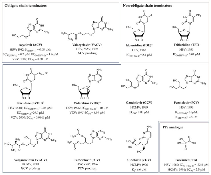
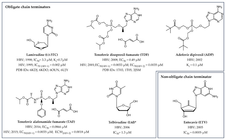
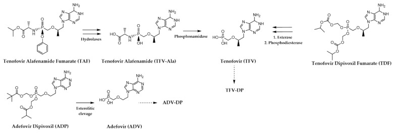
Small molecules that specifically target viral polymerases-crucial enzymes governing viral genome transcription and replication-play a pivotal role in combating viral infections. Presently, approved polymerase inhibitors cover nine human viruses, spanning both DNA and RNA viruses. This review provides a comprehensive analysis of these licensed drugs, encompassing nucleoside/nucleotide inhibitors (NIs), non-nucleoside inhibitors (NNIs), and mutagenic agents. For each compound, we describe the specific targeted virus and related polymerase enzyme, the mechanism of action, and the relevant bioactivity data. This wealth of information serves as a valuable resource for researchers actively engaged in antiviral drug discovery efforts, offering a complete overview of established strategies as well as insights for shaping the development of next-generation antiviral therapeutics.

Keywords: FDA; drugs; inhibitors; small molecules; viral polymerase; viruses.


Conflict of interest statement

The authors declare no conflicts of interest.

Figures

Figure 1
Figure 1
Two-metal ion catalytic mechanism employed by both DNA and RNA polymerases. The primer-dependent mechanism is depicted as an illustrative example (PDB ID 5TXN).
Figure 2
Figure 2
Mechanism of action of obligate chain terminators. (A) Cellular bioactivation of the nucleoside-based (−)-FTC from the non-phosphorylated to the active form. MP, DP, and TP stand for mono-phosphorylated, di-phosphorylated, and tri-phosphorylated, respectively. (B) Simplified 2D representation of the integration of (−)-FTC-TP into the nascent strand. (C) Three-dimensional (3D) representation of (−)-FTC (in cyan) incorporated into the nascent strand (in purple). This image was created using the crystal structure of HIV type 1 (HIV-1) RT in complex with both dsDNA and (−)-FTC (PDB ID: 6WPH). Template strand is in green, whereas yellow dotted lines indicate hydrogen-bond interactions.
Figure 3
Figure 3
Mechanism of action of non-obligate chain terminators. (A) Cellular bioactivation of the prodrug RDV to the TP active form. (B) Simplified 2D representation of the integration of RDV-MP into the nascent strand. (C) A 3D representation of RDV-MP (in cyan) incorporated into the growing strand (in purple). This image was created using the cryogenic electron microscopy (cryo-EM) structure of SARS-CoV-2 RdRp-RNA-inhibitor complex (PDB ID: 7B3C). Template strand is in green, whereas yellow dotted lines indicate intermolecular hydrogen-bond interactions.
Figure 4
Figure 4
Architectural organization of viral polymerases for which experimental structural information is available. Each panel figure contains the respective PDB ID for reference, along with the 3D structure and corresponding PDB sequence of the polymerase domain. Palm, thumb, and fingers subdomains are illustrated in cartoon and colored as blue, magenta, and yellow, respectively. Catalytic Asp residues are represented as red spheres. (A) HSV DdDp. The additional domains Pre-NH2, Exo domain, and NH2-ter domain are showed in white, light blue, and orange, respectively. (B) HCV RdRp. (C) Influenza polymerase (PB1). The PA (green) and PB2 (grey) subunits are represented as surface. (D) RSV RdRp. (E) HIV RT. The p51 and p66 subunits are represented as cartoon and surface, respectively. The RNAse H domain is depicted in green, while the Connection Loop is highlighted in wheat. (F) SARS-CoV-2 RdRp.
Figure 5
Figure 5
The 2D chemical structures of approved small molecule drugs targeting the herpesvirus polymerase activity. a Withdrawn from the market and/or no longer recommended for use in the U.S. (https://www.accessdata.fda.gov/scripts/cder/daf/, accessed on 30 March 2024). b Drugs approved outside U.S.
Figure 6
Figure 6
Bioactivation of prodrugs designed to target herpesvirus polymerases for VACV (A), VGCV (B) and FCV (C).
Figure 7
Figure 7
The 2D chemical structure of approved NRTIs and NtRTIs inhibiting the HBV polymerase activity. (−)-3TC and TDF are also licensed as anti-HIV drugs. a Withdrawn from the market and/or no longer recommended for use in the U.S. (https://www.accessdata.fda.gov/scripts/cder/daf/, accessed on 30 March 2024).
Figure 8
Figure 8
Bioactivation of prodrugs targeting HBV polymerase.
Figure 9
Figure 9
The 2D chemical structure of approved NRTIs and NtRTIs inhibiting the HIV RT polymerase activity as obligate chain terminators. a Withdrawn from the market and/or no longer recommended for use in the U.S. (https://www.accessdata.fda.gov/scripts/cder/daf/, accessed on 30 March 2024).
Figure 10
Figure 10
Bioactivation of prodrug Abacavir (ABC) to CBV-MP.
Figure 11
Figure 11
The 2D chemical structure of approved NNRTIs inhibiting the HIV RT polymerase activity. a Withdrawn from the market and/or no longer recommended for use in the U.S. (https://www.accessdata.fda.gov/scripts/cder/daf/, accessed on 30 March 2024).
Figure 12
Figure 12
(A) Surface representation of the functionally active HIV-1 RT (PDB ID 1DLO [113]). The crucial active site Asp residues are represented by the red ball and stick model, while the surface area representing the allosteric pocket is shaded in cyan. (B) Superimposition of two protein states: the functionally active HIV-RT (wheat, PDB ID 1DLO) and the inactive protein conformation (cyan, PDB ID 4G1Q) when bound to the NNRTI RPV (purple) [102]. For clarity, only residues situated within 4 Å proximity to RPV, along with a RMSD value greater than 2.5 Å between the two protein conformations, are shown.
Figure 13
Figure 13
Patterns of 2D ligand–protein interactions and 3D inhibitor conformations extrapolated from crystal complexes of NNRTIs bound to HIV RT polymerase. The intermolecular interactions were derived using software such as the Schrödinger suite 2021-2 (ligand interaction tool) [115] and LigandScout 4.4 [116] and are illustrated as follows: H-bonds (directed or water-mediated) as purple arrows, hydrophobic interactions as dotted lines with the involved ligand portions highlighted in yellow, and π–π interactions as green lines. The 3D ligand-bound conformation for NVP, DLV, and EFZ resemble a butterfly-like structure, while ETR, RPV, and DOR adopt a “horseshoe” or “U” shape. For each drug, the PDB ID with the best resolution has been used as representative. a Withdrawn from the market and/or no longer recommended for use in the U.S. (https://www.accessdata.fda.gov/scripts/cder/daf/, accessed on 30 March 2024).
Figure 14
Figure 14
The 2D chemical structures of approved small molecule drugs targeting the HCV polymerase. a Withdrawn from the market and/or no longer recommended for use in the U.S. (https://www.accessdata.fda.gov/scripts/cder/daf/, accessed on 30 March 2024). b Drugs approved outside U.S.
Figure 15
Figure 15
Bioactivation of prodrug SOF to the pharmacologically active form GS461203.
Figure 16
Figure 16
HCV NS5B with approved NNIs binding to distinct allosteric sites. Specifically, DSV (green) occupies the PS-I, while BCV (magenta) resides within the TS-I. The experimental position of BCV is derived from the PDB ID 4NLD [130], while the DSV location was generated by SeeSAR 13.0.1 [131] molecular docking using its analogue binder in the PDB ID 4MKB [129] as reference compound. The intermolecular interactions were derived using software such as the Schrödinger suite 2021-2 (ligand interaction tool) [115] and LigandScout 4.4 [116] and are illustrated as follows: H-bonds (directed or water-mediated) as purple arrows, hydrophobic interactions as dotted lines with the involved ligand portions highlighted in yellow, and π–π interactions as green lines Key catalytic residues Asp318, Asp319, and Asp220 are highlighted in red using a ball and stick representation.
Figure 17
Figure 17
(A) The 2D chemical structure of the only approved small molecule targeting the influenza polymerase by multiple mechanisms (e.g., as mutagenic agent). It should be noted that this drug was approved outside the U.S. (B) Bioactivation of prodrug FVP.
Figure 18
Figure 18
The 2D chemical structure of approved small molecules targeting the SARS-CoV-2 polymerase.
Figure 19
Figure 19
Bioactivation of prodrug MOL.
Figure 20
Figure 20
Schematic illustration of the base pairs involving standard nucleobases (A) and the two different tautomeric forms of NHC (B), represented in red, in the RdRp active center.
Figure 21
Figure 21
SARS-CoV-2 RdRp complexed with the two tautomeric forms of NHC. The amino NHC and the imino NHC are base-paired with G (PDB ID: 7OZV) (A) and A (PDB ID: 7OZU) (B), respectively. Hydrogen bonds between NHC and G/A are represented as dashed yellow lines.

References

    1. Li G., Hilgenfeld R., Whitley R., De Clercq E. Therapeutic strategies for COVID-19: Progress and lessons learned. Nat. Rev. Drug Discov. 2023;22:449–475. doi: 10.1038/s41573-023-00672-y. - DOI - PMC - PubMed
    1. Choi K.H. Viral Molecular Machines. Springer; Boston, MA, USA: 2012. Viral polymerases; pp. 267–304. Book Series: Advances in Experimental Medicine and Biology (AEMB, volume 726) - DOI
    1. Pathania S., Rawal R.K., Singh P.K. RdRp (RNA-dependent RNA polymerase): A key target providing anti-virals for the management of various viral diseases. J. Mol. Struct. 2022;1250:131756. doi: 10.1016/j.molstruc.2021.131756. - DOI - PMC - PubMed
    1. Tsai C.H., Lee P.Y., Stollar V., Li M.L. Antiviral therapy targeting viral polymerase. Curr. Pharm. Des. 2006;12:1339–1355. doi: 10.2174/138161206776361156. - DOI - PubMed
    1. De Clercq E., Li G. Approved Antiviral Drugs over the Past 50 Years. Clin. Microbiol. Rev. 2016;29:695–747. doi: 10.1128/CMR.00102-15. - DOI - PMC - PubMed

LinkOut - more resources