. 2024 May 8;16(5):632.
doi: 10.3390/pharmaceutics16050632.

Shape Matters: Impact of Mesoporous Silica Nanoparticle Morphology on Anti-Tumor Efficacy

Affiliations

Shape Matters: Impact of Mesoporous Silica Nanoparticle Morphology on Anti-Tumor Efficacy

Weixiang Fang et al. Pharmaceutics. .

Abstract

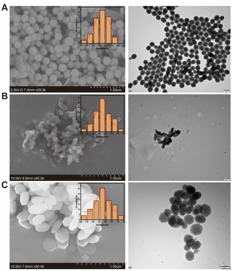
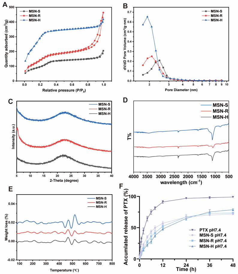
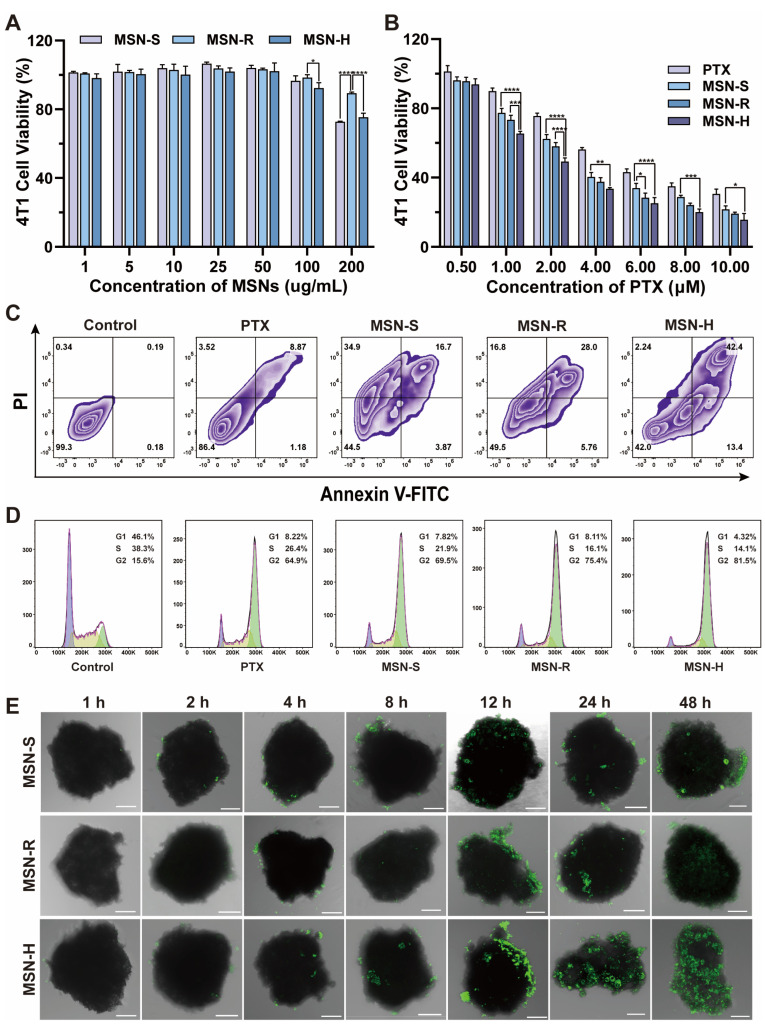
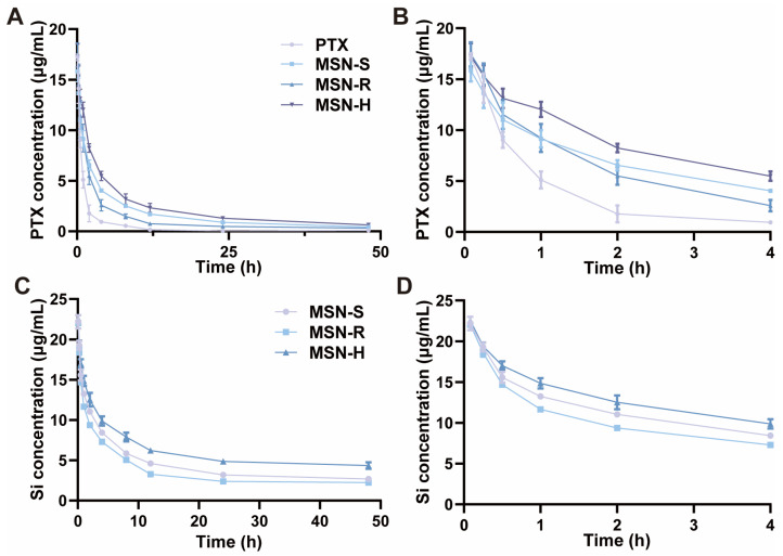
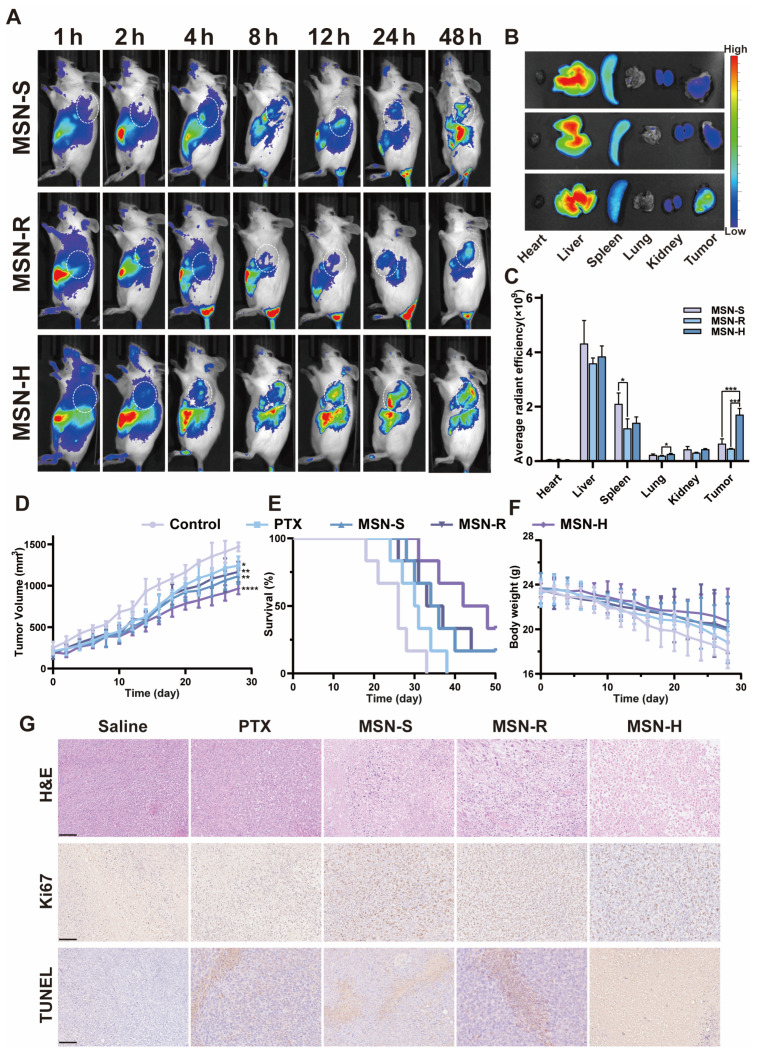
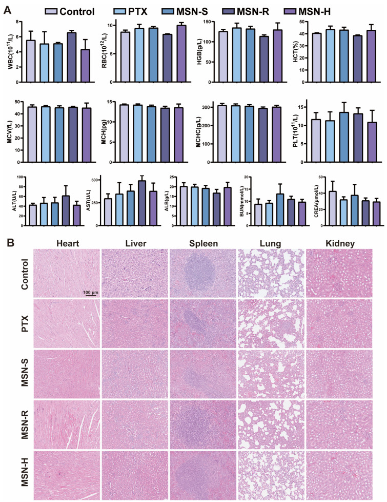
A nanoparticle's shape is a critical determinant of its biological interactions and therapeutic effectiveness. This study investigates the influence of shape on the performance of mesoporous silica nanoparticles (MSNs) in anticancer therapy. MSNs with spherical, rod-like, and hexagonal-plate-like shapes were synthesized, with particle sizes of around 240 nm, and their other surface properties were characterized. The drug loading capacities of the three shapes were controlled to be 47.46%, 49.41%, and 46.65%, respectively. The effects of shape on the release behaviors, cellular uptake mechanisms, and pharmacological behaviors of MSNs were systematically investigated. Through a series of in vitro studies using 4T1 cells and in vivo evaluations in 4T1 tumor-bearing mice, the release kinetics, cellular behaviors, pharmacological effects, circulation profiles, and therapeutic efficacy of MSNs were comprehensively assessed. Notably, hexagonal-plate-shaped MSNs loaded with PTX exhibited a prolonged circulation time (t1/2 = 13.59 ± 0.96 h), which was approximately 1.3 times that of spherical MSNs (t1/2 = 10.16 ± 0.38 h) and 1.5 times that of rod-shaped MSNs (t1/2 = 8.76 ± 1.37 h). This research underscores the significance of nanoparticles' shapes in dictating their biological interactions and therapeutic outcomes, providing valuable insights for the rational design of targeted drug delivery systems in cancer therapy.

Keywords: anticancer therapy; biodistribution; drug delivery; mesoporous silica nanoparticles (MSNs); shape effect.


Conflict of interest statement

The authors declare no conflicts of interest.

Figures

Figure 1
Figure 1
Morphology of MSN with different shapes. Scanning Electron Microscopy (SEM) images and size distribution, as well as Transmission Electron Microscopy (TEM) images of (A) MSN-S, (B) MSN-R, and (C) MSN-H.
Figure 2
Figure 2
Characterization of MSN with different shapes. (A) Nitrogen adsorption–desorption isotherms. (B) Pore size distribution curves. (C) Wide-angle X-ray diffraction (XRD) patterns. (D) Infrared spectra. (E) Thermogravimetric analysis curves. (F) In vitro release profiles of paclitaxel (PTX) from MSN loaded with PTX of different shapes in PBS at 37 °C (n = 3).
Figure 3
Figure 3
Cellular behavior studies. (A) Confocal laser scanning microscopy (CLSM) images and fluorescence analysis (n = 3) of internalization of MSN with different shapes by 4T1 cells. (B) Quantitative analysis by flow cytometry (FCM). (C) Investigation of uptake mechanisms of differently shaped MSN by 4T1 cells. Results of (D) lysosomal distribution, (E) microtubules, (F) Golgi apparatus, (G) endoplasmic reticulum, (H) mitochondria, and (I) fluorescence quantification for MSN-S, MSN-R, and MSN-H. Scale bar: 20 μm (*: p < 0.05, **: p < 0.01, ***: p < 0.005, ****: p < 0.001).
Figure 4
Figure 4
Cellular pharmacology studies. (A) Cell viability of 4T1 cells incubated with (A) empty carriers of different shapes at different concentrations and (B) PTX-loaded MSNs after 48 h (n = 3). (C) Apoptosis of 4T1 cells after 48 h of incubation with PTX-loaded MSNs. (D) Cell cycle analysis of 4T1 cells after 24 h of incubation with PTX-loaded MSNs of different shapes, purple for G1 phase, yellow for S phase, and green for G2 phase. (E) Confocal laser scanning microscopy (CLSM) images of three-dimensional tumor spheroids incubated with MSNs of different shapes for 48 h, scale 100 μm. (*: p < 0.05, **: p < 0.01, ***: p < 0.005, ****: p < 0.001).
Figure 5
Figure 5
In vivo pharmacokinetic curves. SD rats (n = 3) were intravenously injected with MSN-S, MSN-R, MSN-H, and PTX solution at equivalent dose of 5 mg/kg of PTX. (A) Pharmacokinetic curves of PTX in blood. (C) Pharmacokinetic curves of MSN in blood. (B,D) Enlarged views corresponding to 0–4 h portion in (A) and (C), respectively.
Figure 6
Figure 6
In vivo distribution and pharmacodynamics study. (A) In vivo fluorescence distribution images after injection of MSN-S, MSN-R, and MSN-H in tumor-bearing mice, (B) tissue distribution at 48 h, and (C) semi-quantitative fluorescence analysis. (D) Relative tumor volumes, (E) survival rates, and (F) body weights of mice treated with saline, PTX solution, PTX-loaded MSN-S, and MSN-R; (G) H&E, Ki67, and TUNEL staining of tumor tissues. Scale bar: 100 μm. * p < 0.05; ** p < 0.01, ***: p < 0.005, ****: p < 0.001.
Figure 7
Figure 7
In vivo safety study. (A) Hematological and liver–kidney function indicators of mice after different treatment regimens, and (B) H&E staining of major tissues.

Similar articles

Cited by

References

    1. Song X., Zhang Q., Chang M., Ding L., Huang H., Feng W., Xu T., Chen Y. Nanomedicine-Enabled Sonomechanical, Sonopiezoelectric, Sonodynamic, and Sonothermal Therapy. Adv. Mater. 2023;35:e2212259. doi: 10.1002/adma.202212259. - DOI - PubMed
    1. Lepeltier E., Rijo P., Rizzolio F., Popovtzer R., Petrikaite V., Assaraf Y.G., Passirani C. Nanomedicine to Target Multidrug Resistant Tumors. Drug Resist. Updates. 2020;52:100704. doi: 10.1016/j.drup.2020.100704. - DOI - PubMed
    1. Wen P., Ke W., Dirisala A., Toh K., Tanaka M., Li J. Stealth and Pseudo-Stealth Nanocarriers. Adv. Drug Deliv. Rev. 2023;198:114895. doi: 10.1016/j.addr.2023.114895. - DOI - PubMed
    1. Mi P., Cabral H., Kataoka K. Ligand-Installed Nanocarriers toward Precision Therapy. Adv. Mater. 2020;32:e1902604. doi: 10.1002/adma.201902604. - DOI - PubMed
    1. Xu J., Lee S.S., Seo H., Pang L., Jun Y., Zhang R.Y., Zhang Z.Y., Kim P., Lee W., Kron S.J., et al. Quinic Acid-Conjugated Nanoparticles Enhance Drug Delivery to Solid Tumors via Interactions with Endothelial Selectins. Small. 2018;14:e1803601. doi: 10.1002/smll.201803601. - DOI - PMC - PubMed

LinkOut - more resources