. 2024 May 17;16(10):1426.
doi: 10.3390/polym16101426.

Comparison of Printable Biomaterials for Use in Neural Tissue Engineering: An In Vitro Characterization and In Vivo Biocompatibility Assessment

Affiliations

Comparison of Printable Biomaterials for Use in Neural Tissue Engineering: An In Vitro Characterization and In Vivo Biocompatibility Assessment

Miguel Etayo-Escanilla et al. Polymers (Basel). .

Abstract

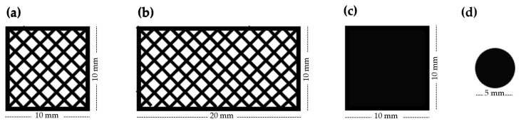
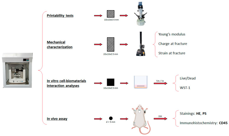
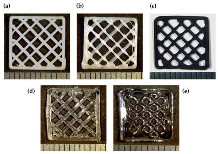
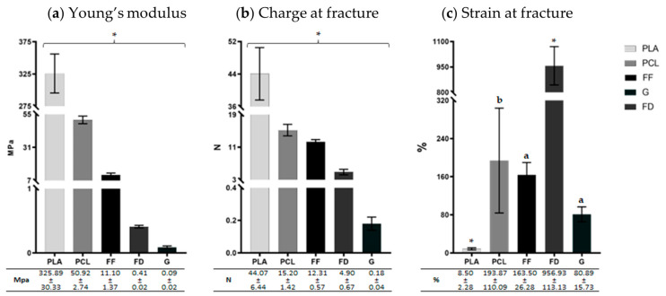
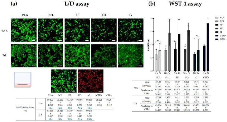
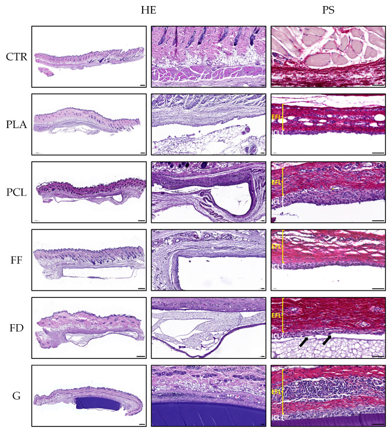
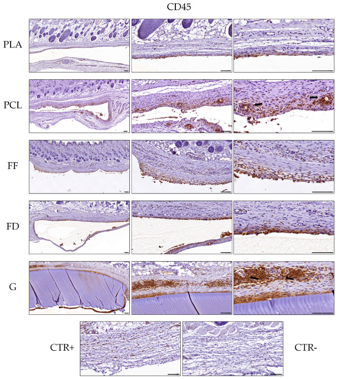
Nervous system traumatic injuries are prevalent in our society, with a significant socioeconomic impact. Due to the highly complex structure of the neural tissue, the treatment of these injuries is still a challenge. Recently, 3D printing has emerged as a promising alternative for producing biomimetic scaffolds, which can lead to the restoration of neural tissue function. The objective of this work was to compare different biomaterials for generating 3D-printed scaffolds for use in neural tissue engineering. For this purpose, four thermoplastic biomaterials, ((polylactic acid) (PLA), polycaprolactone (PCL), Filaflex (FF) (assessed here for the first time for biomedical purposes), and Flexdym (FD)) and gelatin methacrylate (GelMA) hydrogel were subjected to printability and mechanical tests, in vitro cell-biomaterial interaction analyses, and in vivo biocompatibility assessment. The thermoplastics showed superior printing results in terms of resolution and shape fidelity, whereas FD and GelMA revealed great viscoelastic properties. GelMA demonstrated a greater cell viability index after 7 days of in vitro cell culture. Moreover, all groups displayed connective tissue encapsulation, with some inflammatory cells around the scaffolds after 10 days of in vivo implantation. Future studies will determine the usefulness and in vivo therapeutic efficacy of novel neural substitutes based on the use of these 3D-printed scaffolds.

Keywords: 3D printing; biocompatibility; biomaterials; biomechanic; neural tissue engineering; scaffolds.


Conflict of interest statement

The authors declare no conflicts of interest. The funders had no role in the design of the study; in the collection, analyses, or interpretation of data; in the writing of the manuscript; or in the decision to publish the results.

Figures

Figure 1
Figure 1
Design of the scaffolds used for (a) printability tests, (b) mechanical tests, (c) in vitro cell–biomaterial interaction analyses, and (d) in vivo assays.
Figure 2
Figure 2
Scheme of the materials and methods employed in the printability tests, mechanical characterization, the in vitro cell–biomaterial interaction analyses, and the in vivo assay. HE refers to hematoxylin and eosin staining and PS to Picrosirius staining.
Figure 3
Figure 3
Macroscopic images of 3D-printed scaffolds of (a) PLA, (b) PCL, (c) FF, (d) FD, and (e) GelMA. Bright and contrast have been edited to improve their visualization.
Figure 4
Figure 4
Graphic representation of tensile test results of PLA, PCL, FF, FD, and GelMA (G). (a) Young’s modulus (Mpa), (b) charge at fracture (N), and (c) strain at fracture (%). The results corresponding to each mechanical parameter are shown as mean ± standard deviation values. Statistically significant differences were determined with the Mann–Whitney test and represented as follows: ‘*’ indicates statistically significant differences (p < 0.05) between all biomaterials, ‘a’ indicates statistically significant differences (p < 0.05) between all biomaterials except “PCL,” and ‘b’ indicates statistically significant differences (p < 0.05) between all biomaterials except “FF” and “G.”
Figure 5
Figure 5
In vitro biocompatibility tests. (a) Representative panel of L/D assay of SK-N-AS seeded with PLA, PCL, FF, FD, and GelMA (G), and in 2D cultures as technical controls, after 72 h and 7 days of culture. Scale bar = 100 µm. (b) Graphic representation of WST-1 results of PLA, PCL, FF, FD, and GelMA (G). Statistically significant differences were determined with the Mann–Whitney test and are represented as follows: “NS” indicates no significant differences (p ≥ 0.05) between 72 h and 7 days; “2 or 1” indicates the number of biomaterials that obtained a significantly inferior cell viability (Figure 5a) or absorbance value (Figure 5b); and ‘a’ indicates no significant differences (p ≥ 0.05) with the CTR+ group.
Figure 6
Figure 6
Representative panel of the hematoxylin and eosin (HE) and Picrosirius (PS) staining of the in vivo samples of PLA, PCL, FF, FD, GelMA (G), and healthy rat skin tissue (CTR). The PS images distinguish an external fibrotic layer (EFL) and an inner cellular layer (ICL). The black arrows indicate syncytial formations. Scale bar of the first column indicates 500 µm, while the scale bar of the second and third columns indicates 50 µm.
Figure 7
Figure 7
Representative panel of CD45 immunohistochemistry of the in vivo samples of PLA, PCL, FF, FD, GelMA (G), and healthy rat skin tissue (incubated with CD45 antibody (CTR+) and without CD45 antibody (CTR−). The black arrows indicate signs of perivascular infiltration. Scale bar = 100 µm.

Similar articles

Cited by

References

    1. Collaborators G.B.D.N. Global, regional, and national burden of neurological disorders, 1990-2016: A systematic analysis for the Global Burden of Disease Study 2016. Lancet Neurol. 2019;18:459–480. doi: 10.1016/S1474-4422(18)30499-X. - DOI - PMC - PubMed
    1. Singh A., Tetreault L., Kalsi-Ryan S., Nouri A., Fehlings M.G. Global prevalence and incidence of traumatic spinal cord injury. Clin. Epidemiol. 2014;6:309–331. doi: 10.2147/CLEP.S68889. - DOI - PMC - PubMed
    1. Zhang S., Huang M., Zhi J., Wu S., Wang Y., Pei F. Research Hotspots and Trends of Peripheral Nerve Injuries Based on Web of Science From 2017 to 2021: A Bibliometric Analysis. Front. Neurol. 2022;13:872261. doi: 10.3389/fneur.2022.872261. - DOI - PMC - PubMed
    1. Ding W., Hu S., Wang P., Kang H., Peng R., Dong Y., Li F. Spinal Cord Injury: The Global Incidence, Prevalence, and Disability From the Global Burden of Disease Study 2019. Spine. 2022;47:1532–1540. doi: 10.1097/BRS.0000000000004417. - DOI - PMC - PubMed
    1. Carriel V., Garzon I., Alaminos M., Cornelissen M. Histological assessment in peripheral nerve tissue engineering. Neural Regen. Res. 2014;9:1657–1660. doi: 10.4103/1673-5374.141798. - DOI - PMC - PubMed

LinkOut - more resources