

Gender Differences in the Impact of a High-Fat, High-Sugar Diet in Skeletal Muscles of Young Female and Male Mice
- PMID: 38794705
- PMCID: PMC11124085
- DOI: 10.3390/nu16101467
Gender Differences in the Impact of a High-Fat, High-Sugar Diet in Skeletal Muscles of Young Female and Male Mice
Abstract
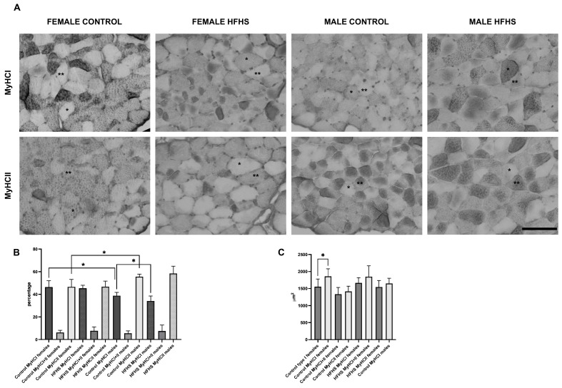
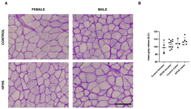
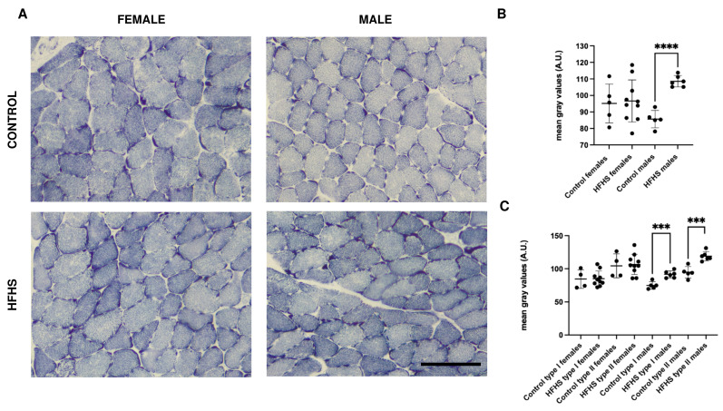
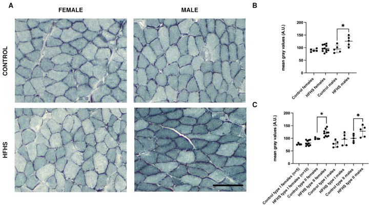
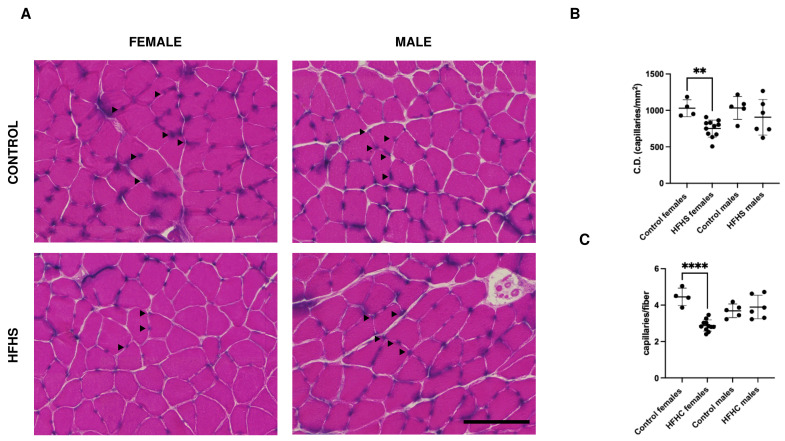
In the context of the increasing number of obese individuals, a major problem is represented by obesity and malnutrition in children. This condition is mainly ascribable to unbalanced diets characterized by high intakes of fat and sugar. Childhood obesity and malnutrition are not only associated with concurrent pathologies but potentially compromise adult life. Considering the strict correlation among systemic metabolism, obesity, and skeletal muscle health, we wanted to study the impact of juvenile malnutrition on the adult skeletal muscle. To this aim, 3-week-old C56BL/6 female and male mice were fed for 20 weeks on a high-fat. high-sugar diet, and their muscles were subjected to a histological evaluation. MyHCs expression, glycogen content, intramyocellular lipids, mitochondrial activity, and capillary density were analyzed on serial sections to obtain the metabolic profile. Our observations indicate that a high-fat, high-sugar diet alters the metabolic profile of skeletal muscles in a sex-dependent way and induces the increase in type II fibers, mitochondrial activity, and lipid content in males, while reducing the capillary density in females. These data highlight the sex-dependent response to nutrition, calling for the development of specific strategies and for a systematic inclusion of female subjects in basic and applied research in this field.
Keywords: Western diet; cell metabolism; fiber plasticity; obesity; skeletal muscle.
Conflict of interest statement
Authors declare no conflict of interests. The funders had no role in the design of the study; in the collection, analyses, or interpretation of data; in the writing of the manuscript; or in the decision to publish the results.
Figures
Similar articles
-
Skeletal muscle and fiber type-specific intramyocellular lipid accumulation in obese mice.Bosn J Basic Med Sci. 2021 Dec 1;21(6):730-738. doi: 10.17305/bjbms.2021.5876. Bosn J Basic Med Sci. 2021. PMID: 34082690 Free PMC article.
-
Impact of 4 weeks of western diet and aerobic exercise training on whole-body phenotype and skeletal muscle mitochondrial respiration in male and female mice.Physiol Rep. 2022 Dec;10(24):e15543. doi: 10.14814/phy2.15543. Physiol Rep. 2022. PMID: 36541261 Free PMC article.
-
Decrement in resting and insulin-stimulated soleus muscle mitochondrial respiration is an early event in diet-induced obesity in mice.Exp Physiol. 2019 Mar;104(3):306-321. doi: 10.1113/EP087317. Epub 2019 Jan 24. Exp Physiol. 2019. PMID: 30578638
-
Fiber-type-specific sensitivities and phenotypic adaptations to dietary fat overload differentially impact fast- versus slow-twitch muscle contractile function in C57BL/6J mice.J Nutr Biochem. 2015 Feb;26(2):155-64. doi: 10.1016/j.jnutbio.2014.09.014. Epub 2014 Oct 25. J Nutr Biochem. 2015. PMID: 25516489
-
Enhanced lipid oxidation and maintenance of muscle insulin sensitivity despite glucose intolerance in a diet-induced obesity mouse model.PLoS One. 2013 Aug 12;8(8):e71747. doi: 10.1371/journal.pone.0071747. eCollection 2013. PLoS One. 2013. PMID: 23951235 Free PMC article.
Cited by
-
Relationship Between Brain Insulin Resistance, Carbohydrate Consumption, and Protein Carbonyls, and the Link Between Peripheral Insulin Resistance, Fat Consumption, and Malondialdehyde.Biomedicines. 2025 Feb 7;13(2):404. doi: 10.3390/biomedicines13020404. Biomedicines. 2025. PMID: 40002817 Free PMC article.
-
Effects of defined voluntary running distances coupled with high-fat diet consumption on the skeletal muscle transcriptome of male mice.Physiol Rep. 2025 Jan;13(2):e70170. doi: 10.14814/phy2.70170. Physiol Rep. 2025. PMID: 39821584 Free PMC article.
References
MeSH terms
Substances
LinkOut - more resources
Full Text Sources
