Observational Study
. 2024 Jul 1;29(4):e575-e583.
doi: 10.4317/medoral.26551.

Insights into incipient oral squamous cell carcinoma: a comprehensive south-american study

Affiliations
Observational Study

Insights into incipient oral squamous cell carcinoma: a comprehensive south-american study

C Saldivia-Siracusa et al. Med Oral Patol Oral Cir Bucal. .

Abstract

Background: To describe demographic and clinicopathological aspects of a South-American cohort of incipient oral squamous cell carcinoma patients.

Material and methods: A cross-sectional, observational study was performed to assess demographic and clinicopathological characteristics of incipient oral squamous cell carcinoma patients from 6 South-American institutions.

Results: One hundred and seven patients within the histopathological spectrum of incipient oral squamous cell carcinoma (in-situ and microinvasive) were included. Fifty-eight (54.2%) patients were men with a mean age of 60.69 years. Forty-nine (45.8%) and thirty-nine (36.5%) patients had history of tobacco and alcohol use, respectively. Clinically, most of the lesions were plaques (82.2%), ≥ 2 cm in extension (72%), affecting the lateral border of the tongue (55.1%), and soft palate (12.1%) with a mixed (white and red) appearance. Eighty-two (76.7%) lesions were predominantly white and 25 (23.3%) predominantly red.

Conclusions: To the best of our knowledge, this is the largest cohort of incipient oral squamous cell carcinoma patients, which raises awareness of clinicians' inspection acuteness by demonstrating the most frequent clinical aspects of this disease, potentially improving oral cancer secondary prevention strategies.


Conflict of interest statement

The authors declare no conflict of interest, financial or otherwise.

Figures

Figure 1
Figure 1
Incipient oral squamous cell carcinoma cases with subtle clinical presentation. a) Homogeneous leukoplakia removed through excisional biopsy, resulting on a microinvasive OSCC; b) Erythroleukoplakia of the soft palate treated through excisional biopsy, showing in-situ OSCC; c) Mixed leukoerythroplakia with a diagnosis of in-situ OSCC; d) Microinvasive OSCC noticed as a small ulcer without indurated borders, affecting marginal gingiva; e) Small mixed lesion resulting in an in-situ OSCC: f) Nodular leukoplakia of lateral border of tongue with diagnosis of in-situ OSCC; g) Non homogeneous plaque situated on retromolar trigone, diagnosed as in-situ OSCC. Due to posterior localization and smaller size, the identification of this lesion could easily be overlooked by the patient or during a careless visual examination; h) Microinvasive OSCC presenting as a non-homogeneous speckled leukoplakia comprising the right side of the soft palate.
Figure 2
Figure 2
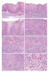
Histopathological spectrum of OSCCi cases (Hematoxylin and eosin). a) Severe epithelial dysplasia/in-situ carcinoma of the buccal mucosa showing atypia involving total thickness of the epithelium without basal membrane breach (10X); b) Leukoerytrhoplakia of the lateral border of tongue diagnosed as severe epithelial dysplasia/in-situ carcinoma. Lichenoid immune response is present in the subjacent connective tissue (10X); c) Non-homogeneous leukoplakia diagnosed as a severe epithelial dysplasia/in-situ carcinoma exhibiting bulbous rete processes and architectural disorganization (20X); d) Severe epithelial dysplasia/in-situ displaying architectural and cytologic atypia throughout total epithelium thickness (20X); e) OSCCmi showing keratinizing epithelial islands invading the subjacent connective tissue below 5 mm depth (20X); f) Basal membrane breach characterized by atypical basal cells showing loss of cohesion, as well as discrete small epithelial islands surrounded by chronic immune response (20X); g) Mixed lesion showing intense atypia and discrete microinvasive foci confined to the lamina propria (20X); h) Scarce epithelial cells showing cellular and nuclear atypia, focally branching outside the epithelium in a OSCCmi (40X).

References

    1. Bouvard V, Nethan ST, Singh D, Warnakulasuriya S, Mehrotra R, Chaturvedi AK. IARC Perspective on Oral Cancer Prevention. N Engl J Med. 2022;387:1999–2005. - PubMed
    1. Louredo BV, Vargas PA, Pérez-de-Oliveira ME, Lopes MA, Kowalski LP, Curado MP. Epidemiology and survival outcomes of lip, oral cavity, and oropharyngeal squamous cell carcinoma in a southeast Brazilian population. Med Oral Patol Oral Cir Bucal. 2022;27:e274–84. - PMC - PubMed
    1. Seoane J, Takkouche B, Varela-Centelles P, Tomás I, Seoane-Romero JM. Impact of delay in diagnosis on survival to head and neck carcinomas: a systematic review with meta-analysis. Clin Otolaryngol. 2012;37:99–106. - PubMed
    1. González-Moles MÁ, Aguilar-Ruiz M, Ramos-García P. Challenges in the Early Diagnosis of Oral Cancer, Evidence Gaps and Strategies for Improvement: A Scoping Review of Systematic Reviews. Cancers (Basel) 2022;14:4967. - PMC - PubMed
    1. Almangush A, Bello IO, Coletta RD, Mäkitie AA, Mäkinen LK, Kauppila JH. For early-stage oral tongue cancer, depth of invasion and worst pattern of invasion are the strongest pathological predictors for locoregional recurrence and mortality. Virchows Arch. 2015;467:39–46. - PubMed

Publication types

LinkOut - more resources