

Liver fibrosis is associated with left ventricular remodeling: insight into the liver-heart axis
- PMID: 38795131
- PMCID: PMC11519090
- DOI: 10.1007/s00330-024-10798-1
Liver fibrosis is associated with left ventricular remodeling: insight into the liver-heart axis
Abstract
Objective: In nonalcoholic fatty liver disease (NAFLD), liver fibrosis is the strongest predictor of adverse outcomes. We sought to investigate the relationship between liver fibrosis and cardiac remodeling in participants from the general population using magnetic resonance imaging (MRI), as well as explore potential mechanistic pathways by analyzing circulating cardiovascular biomarkers.
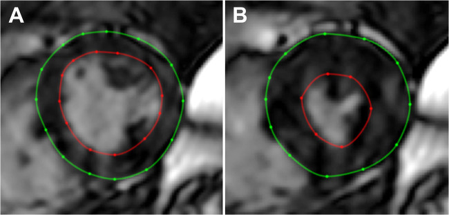
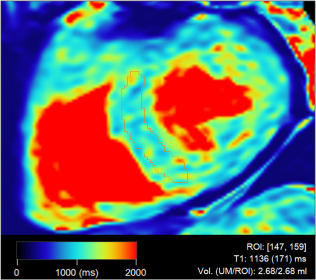
Methods: In this cross-sectional study, we prospectively included participants with type 2 diabetes and individually matched controls from the SCAPIS (Swedish CArdioPulmonary bioImage Study) cohort in Linköping, Sweden. Between November 2017 and July 2018, participants underwent MRI at 1.5 Tesla for quantification of liver proton density fat fraction (spectroscopy), liver fibrosis (stiffness from elastography), left ventricular (LV) structure and function, as well as myocardial native T1 mapping. We analyzed 278 circulating cardiovascular biomarkers using a Bayesian statistical approach.
Results: In total, 92 participants were enrolled (mean age 59.5 ± 4.6 years, 32 women). The mean liver stiffness was 2.1 ± 0.4 kPa. 53 participants displayed hepatic steatosis. LV concentricity increased across quartiles of liver stiffness. Neither liver fat nor liver stiffness displayed any relationships to myocardial tissue characteristics (native T1). In a regression analysis, liver stiffness was related to increased LV concentricity. This association was independent of diabetes and liver fat (Beta = 0.26, p = 0.0053), but was attenuated (Beta = 0.17, p = 0.077) when also adjusting for circulating levels of interleukin-1 receptor type 2.
Conclusion: MRI reveals that liver fibrosis is associated to structural LV remodeling, in terms of increased concentricity, in participants from the general population. This relationship could involve the interleukin-1 signaling.
Clinical relevance statement: Liver fibrosis may be considered a cardiovascular risk factor in patients without cirrhosis. Further research on the mechanisms that link liver fibrosis to left ventricular concentricity may reveal potential therapeutic targets in patients with non-alcoholic fatty liver disease (NAFLD).
Key points: Previously, studies on liver fibrosis and cardiac remodeling have focused on advanced stages of liver fibrosis. Liver fibrosis is associated with left ventricular (LV) concentricity and may relate to interleukin-1 receptor type 2. Interleukin-1 signaling is a potential mechanistic interlink between early liver fibrosis and LV remodeling.
Keywords: Elastography; Interleukin-1; Magnetic Resonance; Non-alcoholic fatty liver disease; Type 2 diabetes.
© 2024. The Author(s).
Conflict of interest statement
MW is a consultant for SyntheticMR AB and holds stock in SyntheticMR AB. The remaining authors of this manuscript declare no relationships with any companies whose products or services may be related to the subject matter of the article.
Figures


References
-
- Younossi ZM, Golabi P, de Avila L et al (2019) The global epidemiology of NAFLD and NASH in patients with type 2 diabetes: a systematic review and meta-analysis. J Hepatol 71:793–801. 10.1016/j.jhep.2019.06.021 - PubMed
-
- Mantovani A, Byrne CD, Benfari G, Bonapace S, Simon TG, Targher G (2022) Risk of heart failure in patients with nonalcoholic fatty liver disease: JACC review topic of the week. J Am Coll Cardiol 79:180–191. 10.1016/j.jacc.2021.11.007 - PubMed
-
- Valbusa F, Agnoletti D, Scala L et al (2018) Non-alcoholic fatty liver disease and increased risk of all-cause mortality in elderly patients admitted for acute heart failure. Int J Cardiol 265:162–168. 10.1016/j.ijcard.2018.04.129 - PubMed
MeSH terms
Substances
LinkOut - more resources
Full Text Sources
Medical
