

Trans-eyelid distribution of epinastine to the conjunctiva following eyelid application in rabbits
- PMID: 38795193
- PMCID: PMC11420250
- DOI: 10.1007/s10384-024-01070-6
Trans-eyelid distribution of epinastine to the conjunctiva following eyelid application in rabbits
Abstract
Purpose: To reveal the penetration of epinastine, an anti-allergic ophthalmic agent, into the eyelid and its distribution to the conjunctiva after administration of a cream formulation on rabbit eyelid skin.
Study design: Experimental study.
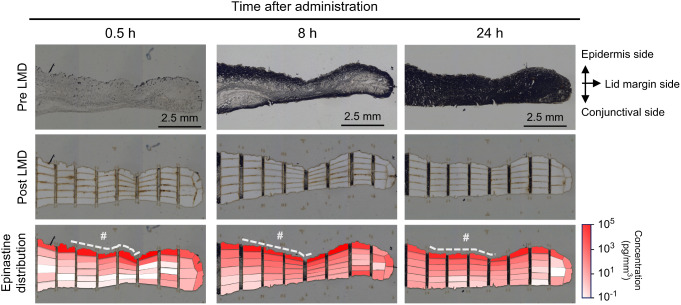
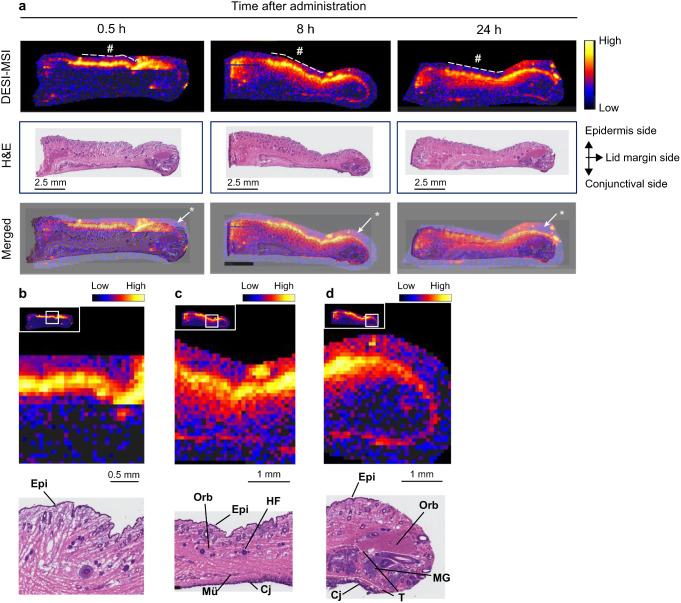
Methods: Rabbits were treated with 0.5% epinastine cream on hair-shaved eyelids, followed by preparation of eyelid tissue slices to determine spatial tissue distribution of epinastine by liquid chromatography-tandem mass spectrometry (LC-MS/MS) quantification using laser-microdissected tissues and desorption electrospray ionization mass spectrometry imaging (DESI-MSI). In addition, following either eyelid application of 0.5% epinastine cream or ocular instillation of 0.1% epinastine eye drops, concentration-time profiles of epinastine in the palpebral conjunctiva and bulbar conjunctiva were determined using LC-MS/MS.
Results: Laser microdissection coupled with LC-MS/MS analysis detected high concentrations of epinastine around the outermost layer of the eyelid at 0.5 h post-administration that gradually diffused deeper into the eyelid and was distributed in the conjunctival layer at 8 and 24 h post-administration. Similar time-dependent drug distribution was observed in high-spatial-resolution images obtained using DESI-MSI. Epinastine concentrations in the conjunctival tissues peaked at 4-8 h after administration of 0.5% epinastine cream and then decreased slowly over 72 h post-administration. In contrast, epinastine concentrations peaked quickly and decreased sharply after epinastine eye drop administration.
Conclusion: After the application of epinastine cream to the eyelid skin, epinastine gradually permeated the eyelid. The compound was retained in the conjunctiva for 8-24 h post-administration, indicating that epinastine cream is a promising long-acting formulation for treating allergic conjunctivitis.
Keywords: Allergic conjunctivitis; Epinastine; Eyelid; Imaging; Ocular distribution.
© 2024. The Author(s).
Conflict of interest statement
T. Mochizuki, Employee (Santen); T. Hata, Employee (Santen); N. Mori, Employee (Santen); T. Yamazaki, Employee (Santen); T. Noto, Employee (Santen); H. Mano, Employee (Santen).
Figures

References
-
- Abelson MB, Shetty S, Korchak M, Butrus SI, Smith LM. Advances in pharmacotherapy for allergic conjunctivitis. Expert Opin Pharmacother. 2015;16:1219–31. - PubMed
-
- Chrai SS, Patton TF, Mehta A, Robinson JR. Lacrimal and instilled fluid dynamics in rabbit eyes. J Pharm Sci. 1973;62:1112–21. - PubMed
-
- Pahuja P, Arora S, Pawar P. Ocular drug delivery system: a reference to natural polymers. Expert Opin Drug Deliv. 2012;9:837–61. - PubMed
-
- Leonardi A, Silva D, Formigo DP, Bozkurt B, Sharma V, Allegri P, et al. Management of ocular allergy. Allergy. 2019;74:1611–30. - PubMed
MeSH terms
Substances
LinkOut - more resources
Full Text Sources
