. 2024 Jun 21;5(2):103087.
doi: 10.1016/j.xpro.2024.103087. Epub 2024 May 23.

Protocol for the development of mRNA lipid nanoparticle vaccines and analysis of immunization efficiency in mice

Affiliations

Protocol for the development of mRNA lipid nanoparticle vaccines and analysis of immunization efficiency in mice

Neha Karekar et al. STAR Protoc. .

Abstract

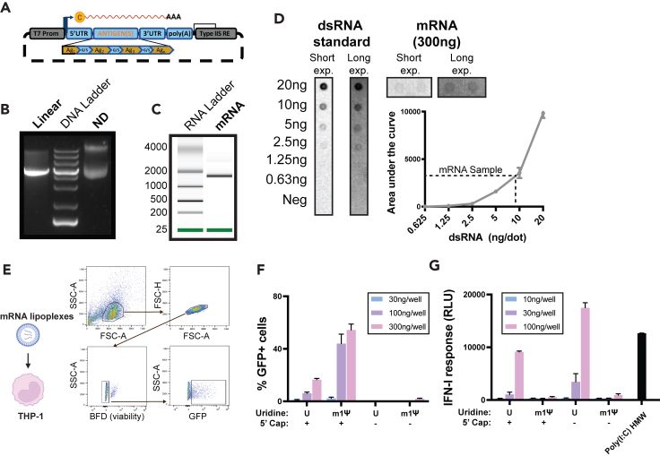
Here, we present a protocol for the development of mRNA-loaded lipid nanoparticle (LNP) vaccines for target antigen sequences of interest. We describe key steps required to design and synthesize mRNA constructs, their LNP encapsulation, and mouse immunization. We then detail quality control assays to determine RNA purity, guidelines to measure RNA immunogenicity using in vitro reporter systems, and a technique to evaluate antigen-specific T cell responses following immunization.

Keywords: biotechnology and bioengineering; cancer; immunology.


Conflict of interest statement

Declaration of interests N.B. serves as an advisor/board member for Apricity, BioNTech, Boehringer Ingelheim, BreakBio, Carisma Therapeutics, CureVac, Genotwin, Gilead, Novartis, PrimeVax, ROME Therapeutics, Tempest Therapeutics, and Rubius Therapeutics and as a consultant for Genentech. N.B. provides research for support for DC Prime, Dragonfly Therapeutics, Inc., Harbor Biomed Sciences, and Regeneron Pharmaceuticals, Inc. N.B. serves on the Scientific Advisory Council/Board of the Cancer Research Institute, Duke University CHAVD, MD Anderson Cancer Center, Parker Institute for Cancer Immunotherapy, and the American Association for Cancer Research. N.B. serves on the grants/research support council for the Cancer Research Institute, Melanoma Research Alliance, Leukemia & Lymphoma Society, Pershing Square Sohn Cancer Research Alliance, and Stand Up to Cancer.

Figures

None
Graphical abstract
Figure 1
Figure 1
Characterization of mRNA purity and immunogenicity (A) Schematic depiction of the plasmid DNA sequence. (B) Gel electrophoresis of linearized plasmid compared to non-digested plasmid (ND). (C) Bioanalyzer electrophoresis mRNA (D). J2 Dot-blot assay quantifying dsRNA quantity in IVT samples using a dsRNA standard (E). Flow cytometry gating strategy quantifying GFP+ transduced THP-1 cells. (F) Comparison of percent GFP translation in THP-1 cells transfected with capped or uncapped mRNAs synthesized using unmodified uridine or modified N1-Methyl-pseudouridine nucleotides. (G) Type-I interferon response measure after transfection of THP-1 reporter cells with same RNAs as in (F).
Figure 2
Figure 2
Determination of the physicochemical and stability properties of fresh and frozen mRNA-LNPs (A) Size distribution of three independent mRNA-LNPs formulations as measured by DLS. (B) Mean hydrodynamic diameter and polydispersity index (PDI) measured via DLS. Results are displayed as mean ± SD. (C) Encapsulation efficiency of mRNA within LNPs of three independent formulations. Results are displayed as mean ± SD. (D) Mean diameter and PDI of mRNA-LNPs after storage at 4C for 1, 2, 3, or 4 weeks. (E) Zeta-potential of the mRNA-LNP after storage at 4C for 1, 2, 3, or 4 weeks. (F) Comparison of mRNA encapsulation efficiency after 1 month of mRNA-LNP storage at 4°C. (G) Size distribution of fresh vs. frozen mRNA-LNPs. (H) Mean diameter and PDI of fresh and frozen LNPs following thawing. (I) Encapsulation efficiency of mRNA-LNP after thawing.
Figure 3
Figure 3
B16F10 and BMDC antigen presentation assay following stimulation with RNA-lipoplexes and RNA-LNP formulations (A) The principle underlying the antigen (SIINFEKL) presentation assay. (B) Flow cytometry gating strategy to quantify antigen presenting (H2-Kb/SIINFEKL+) B16F10 cells. (C) % H2-Kb /SIINFEKL+ cells and (D). Mean fluorescence intensity of H2-Kb /SIINFEKL in B16F10 transduced with fresh or previously frozen mRNA-LNPs. (E) Flow cytometry gating strategy to quantify antigen presenting (H2-Kb/SIINFEKL+) BMDCs. (F) % H2-Kb/SIINFEKL+ cells and (G). Mean fluorescence intensity of H2-Kb /SIINFEKL in BMDCs transduced with fresh or previously frozen mRNA-LNPs.
Figure 4
Figure 4
Induction of antigen-specific T cells and anti-tumor immune responses following mRNA-LNP vaccination (A) Schematic of the prime-boost vaccination assay. (B) Flow cytometry gating strategy to identify antigen-specific CD8+ T cells in peripheral blood mononuclear cells (PBMCs). (C) Percentage of antigen-specific CD8+ T cells among total CD8+ T cells in PBMCs following vaccination with mRNA-LNP. The antigen-specific response to 2 antigens encoded by the vaccines (SIINFEKL/OVA and HSF2) are shown. FMO is a negative control of staining. (D) Flow cytometry gating strategy to identify antigen-specific CD8+ T cells in spleen. (E) Percentage of antigen-specific CD8+ T cells among total CD8+ T cells in spleen following vaccination. (F) Mean tumor growth post engraftment (G). Tumor weight at study endpoint. The significance of the mean tumor weight difference between the control and vaccinated groups is determined by student’s unpaired T-test (p = 0.01).

References

    1. Fang E., Liu X., Li M., Zhang Z., Song L., Zhu B., Wu X., Liu J., Zhao D., Li Y. Advances in COVID-19 mRNA vaccine development. Signal Transduct. Target. Ther. 2022;7:94. doi: 10.1038/s41392-022-00950-y. - DOI - PMC - PubMed
    1. Pingoud A., Jeltsch A. Structure and function of type II restriction endonucleases. Nucleic Acids Res. 2001;29:3705–3727. doi: 10.1093/nar/29.18.3705. - DOI - PMC - PubMed
    1. Mu X., Hur S. Immunogenicity of in vitro-Transcribed RNA. Acc. Chem. Res. 2021;54:4012–4023. doi: 10.1021/acs.accounts.1c00521. - DOI - PMC - PubMed
    1. Baiersdörfer M., Boros G., Muramatsu H., Mahiny A., Vlatkovic I., Sahin U., Karikó K. A Facile Method for the Removal of dsRNA Contaminant from In Vitro-Transcribed mRNA. Mol. Ther. Nucleic Acids. 2019;15:26–35. doi: 10.1016/j.omtn.2019.02.018. - DOI - PMC - PubMed
    1. Vabret N., Blander, J.M. Sensing microbial RNA in the cytosol. Front. Immunol. 2013;4:468. doi: 10.3389/fimmu.2013.00468. - DOI - PMC - PubMed