Review
. 2024 Jun 19;112(12):1911-1929.
doi: 10.1016/j.neuron.2024.05.001. Epub 2024 May 24.

Neurobiological basis of stress resilience

Affiliations
Review

Neurobiological basis of stress resilience

Eric J Nestler et al. Neuron. .

Abstract

A majority of humans faced with severe stress maintain normal physiological and behavioral function, a process referred to as resilience. Such stress resilience has been modeled in laboratory animals and, over the past 15 years, has transformed our understanding of stress responses and how to approach the treatment of human stress disorders such as depression, post-traumatic stress disorder (PTSD), and anxiety disorders. Work in rodents has demonstrated that resilience to chronic stress is an active process that involves much more than simply avoiding the deleterious effects of the stress. Rather, resilience is mediated largely by the induction of adaptations that are associated uniquely with resilience. Such mechanisms of natural resilience in rodents are being characterized at the molecular, cellular, and circuit levels, with an increasing number being validated in human investigations. Such discoveries raise the novel possibility that treatments for human stress disorders, in addition to being geared toward reversing the damaging effects of stress, can also be based on inducing mechanisms of natural resilience in individuals who are inherently more susceptible. This review provides a progress report on this evolving field.

Keywords: adaptation; blood-brain barrier; chronic social defeat stress; coping; depression; nucleus accumbens; post-traumatic stress disorder; prefrontal cortex; stress susceptibility; transcriptomics.


Conflict of interest statement

Declaration of interests The authors declare no competing interests

Figures

Figure 1.
Figure 1.. Resilience and susceptibility circuits.
Left: Cartoon of sagittal brain slice depicting active changes to brain circuits that control reward/valence, attention and cognitive control, or sensory processing that are unique to resilient (RES) mice relative to control (CTRL) and susceptible (SUS) mice after chronic stress. Such active changes include increased or decreased circuit activity or the maintenance of normal activity via a RES-unique mechanism that overcomes a SUS-associated pathological change. Right: Cartoon of sagittal brain slice depicting active changes to brain circuits that control reward/valence, threat/salience, or attention and cognitive control that are unique to SUS mice relative to CTRL and RES mice after chronic stress. VTA, ventral tegmental area; NAc, nucleus accumbens; PFC, prefrontal cortex; LC, locus coeruleus; HIPP, hippocampus; LS, lateral septum; AMY, amygdala; VS, ventral subiculum.
Figure 2.
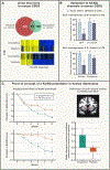
Figure 2.. KCNQ potentiators: Example of advancing a mechanism of stress resilience in mice to novel therapeutics for human depression.
Krishnan et al. (2007) used gene expression microarrays to identify changes in gene expression that occur in the ventral tegmental area (VTA) after chronic social defeat stress in mice that are more susceptible to the stress vs. those that are more resilient (see Box 1 figure). Top heatmap shows genes that are significantly altered (yellow, up; blue, down) in the VTA of susceptible mice and how those same genes are affected in resilience, whereas the bottom heatmap shows the opposite. Among genes induced uniquely in the more resilient VTA are several subunits of KCNQ K+ (Kv7) channels. The authors also showed that viral-mediated overexpression of K+ channels in VTA neurons enhanced behavioral resilience, with a similar effect seen upon systemic administration of ezogabine, a KCNQ channel potentiator that penetrates the blood-brain barrier. These findings in mice provided the rationale for an open-label study of ezogabine in depressed humans, which in turn justified a double-blind, placebo-controlled study which demonstrated ezogabine’s antidepressant efficacy and concomitant correction of functional magnetic resonance deficits in the nucleus accumbens, which along with the VTA comprises the brain’s reward circuitry.
Figure 3.
Figure 3.. Example of the discovery and validation of a novel mechanism of stress resilience in mice.
RNAseq data of multiple limbic brain regions of mice after chronic social defeat stress were analyzed by gene co-expression network analysis to identify gene modules that associate with behavioral resilience. The top-ranked resilience module in medial prefrontal cortex (mPFC) – arbitrarily given the name “pink module” – is enriched in genes related to signal transduction and protein kinase regulation, in contrast to the next two top-ranked modules that are enriched in genes related to synaptic transmission or nerve ensheathment. The panel at the right shows the gene constituents of the pink module, with the strongest hub gene being Zfp189, a putative transcription factor which had not previously been studied in brain. Viral-mediated overexpression of ZFP189 in mPFC neurons triggered robust regulation of a large percentage of pink module genes, with an order of magnitude fewer genes affected in all other modules—thus providing critical empirical validation of the bioinformatic predictions. Such viral overexpression of ZFP189 also increased behavioral resilience in male and female mice subjected to several types of stress (see Ref 112 for details). MDC, module differential connectivity.
Figure 4.
Figure 4.. Active resilience mechanisms that promote blood-brain barrier health.
A. Schematic showing increases in permissive histone acetylation and decreases in repressive histone methylation at the claudin 5 (Cldn5) promoter in nucleus accumbens (NAc) of resilient mice. B. Venn diagram showing overlap between the NAc endothelial transcriptome in susceptible (SUS), resilient (RES), and control CTRL mice following 10 days of CSDS (upper left). Resilient mice exhibit unique transcriptional changes (see Ref 139 for details). C. Schematic summarizing differences in blood-brain barrier (BBB) and immune infiltration in SUS and RES mice compared to CTRL mice. SUS mice exhibit increases in circulating monocytes that traffic to the neurovasculature and release cytokines and matrix metalloproteinases (MMPs) in response to stress that infiltrate the brain parenchyma to regulate neural activity. RES mice have fewer circulating monocytes – that largely remain in the periphery due to a healthy BBB – and release less cytokines and MMPs in response to stress. Panel B is modified with permission from R. Kalisch et al., Neurobiology and systems biology of stress resilience. Physiol Rev, in press.
Box 1 Figure
Box 1 Figure
The figure offers a composite view of “susceptibility” and “resilience” in the CSDS procedure. Historically, this dichotomous designation is based on a rapid social interaction test after 10 days of CSDS in male mice. So-designated susceptible mice, but not resilient mice, also show a deficit in sucrose preference, whereas both groups show a deficit in the elevated plus maze. However, the individual social interaction data show a continuous distribution and, since the original study, several behavioral outcome measures have been analyzed with this mind. Some measures correlate – to different degrees of strength – with social interaction, whereas others do not. Examples of the former are sucrose preference, reversal learning, and some measures from Restaurant Row. An example of the latter is another measure from Restaurant Row. Note social interaction time is shown in the top panel, while the bottom panels use a social interaction ratio (time spent in the presence vs. absence of a social target).

References

    1. Gillespie CF, Bradley B, Mercer K, Smith AK, Conneely K, Gapen M, Weiss T, Schwartz AC, Cubells JF, Ressler KJ. Trauma exposure and stress-related disorders in inner city primary care patients. Gen Hosp Psychiatry. 2009. Nov-Dec;31(6):505–14. - PMC - PubMed
    1. Charney DS. The psychobiology of resilience and vulnerability to anxiety disorders: implications for prevention and treatment. Dialogues Clin Neurosci 2003. Sep;5(3):207–21. - PMC - PubMed
    1. Comtesse H, Powell S, Soldo A, Hagl M, Rosner R. Long-term psychological distress of Bosnian war survivors: an 11-year follow-up of former displaced persons, returnees, and stayers. BMC Psychiatry. 2019. Jan 3;19(1):1. - PMC - PubMed
    1. Andreassen OA, Hindley GFL, Frei O, Smeland OB. New insights from the last decade of research in psychiatric genetics: discoveries, challenges and clinical implications. World Psychiatry. 2023. Feb;22(1):4–24. - PMC - PubMed
    1. Turner CA, Khalil H, Murphy-Weinberg V, Hagenauer MH, Gates L, Tang Y, Weinberg L, Grysko R, Floran-Garduno L, Dokas T, Samaniego C, Zhao Z, Fang Y, Sen S, Lopez JF, Watson SJ Jr, Akil H. The impact of COVID-19 on a college freshman sample reveals genetic and nongenetic forms of susceptibility and resilience to stress. Proc Natl Acad Sci U S A 2023. Dec 5;120(49):e2305779120. - PMC - PubMed

MeSH terms

LinkOut - more resources