. 2024 Jul;131(1):77-89.
doi: 10.1038/s41416-024-02689-5. Epub 2024 May 25.

Oncofetal SNRPE promotes HCC tumorigenesis by regulating the FGFR4 expression through alternative splicing

Affiliations

Oncofetal SNRPE promotes HCC tumorigenesis by regulating the FGFR4 expression through alternative splicing

Qipeng Wu et al. Br J Cancer. 2024 Jul.

Abstract

Background: Due to insufficient knowledge about key molecular events, Hepatocellular carcinoma (HCC) lacks effective treatment targets. Spliceosome-related genes were significantly altered in HCC. Oncofetal proteins are ideal tumor therapeutic targets. Screening of differentially expressed Spliceosome-related oncofetal protein in embryonic liver development and HCC helps discover effective therapeutic targets for HCC.

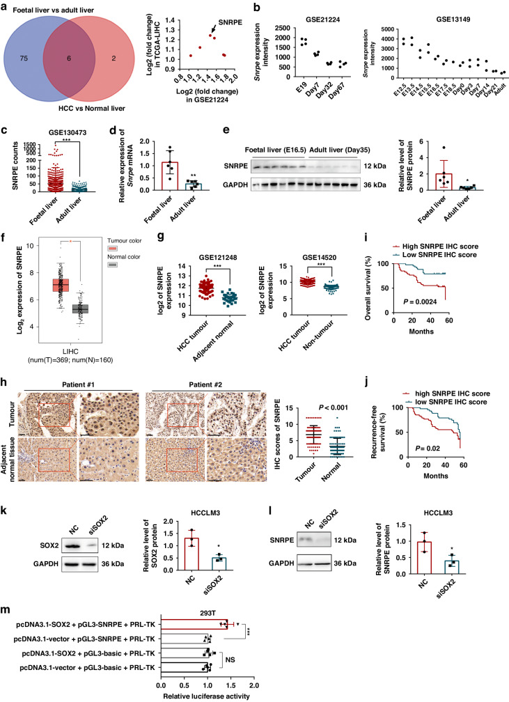
Methods: Differentially expressed spliceosome genes were analysis in fetal liver and HCC through bioinformatics analysis. Small nuclear ribonucleoprotein polypeptide E (SNRPE) expression was detected in fetal liver, adult liver and HCC tissues. The role of SNRPE in HCC was performed multiple assays in vitro and in vivo. SNRPE-regulated alternative splicing was recognized by RNA-Seq and confirmed by multiple assays.

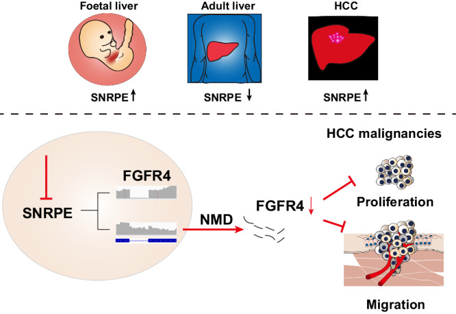
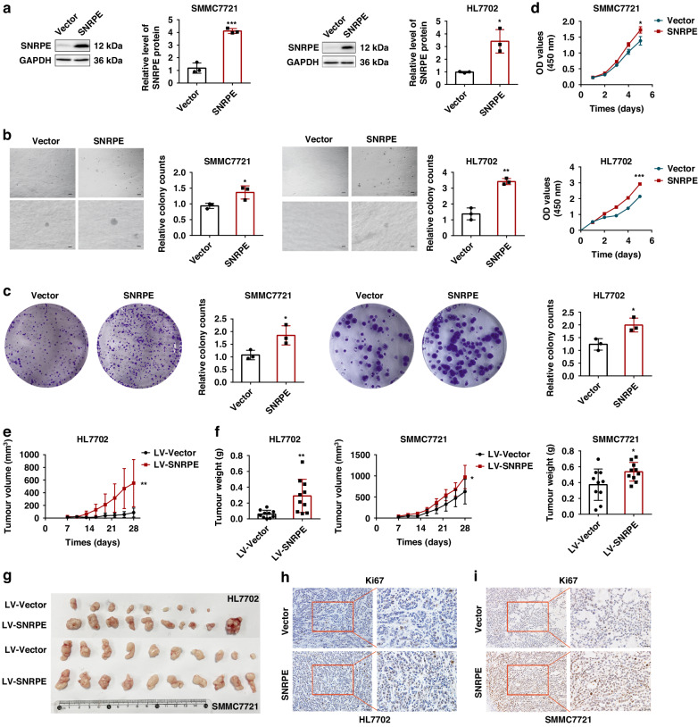
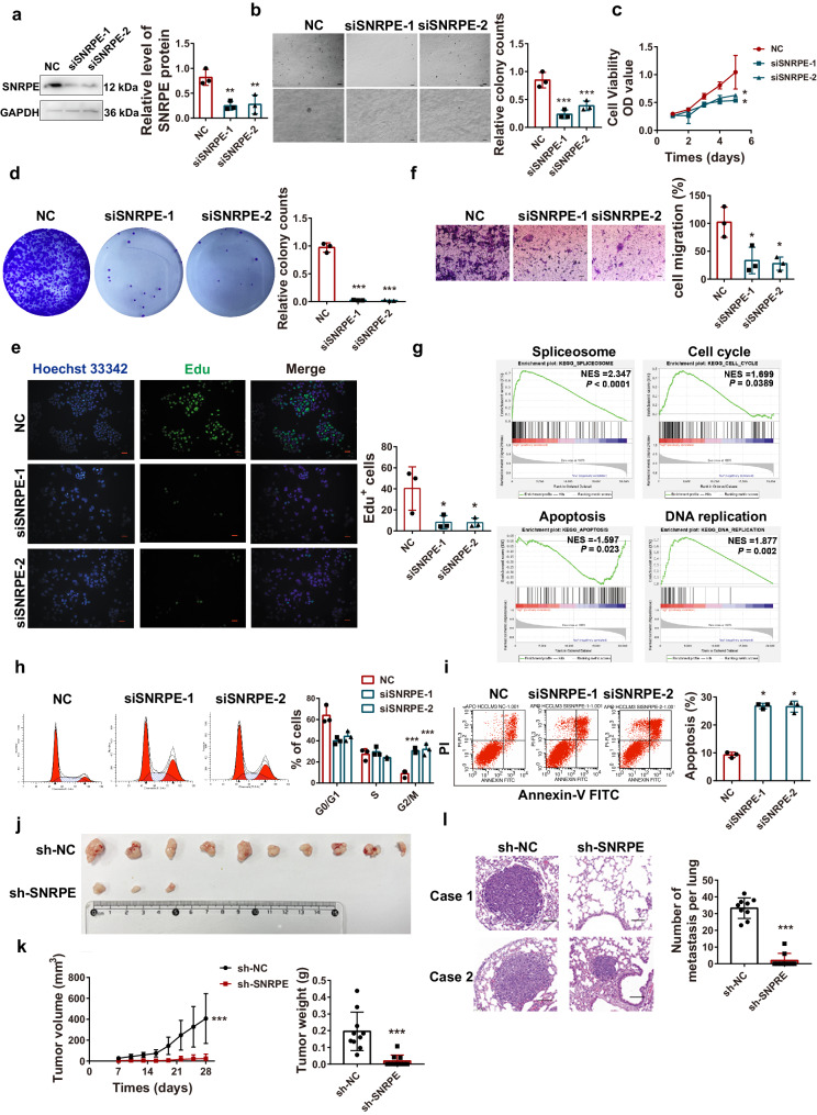
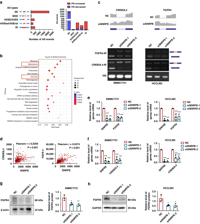
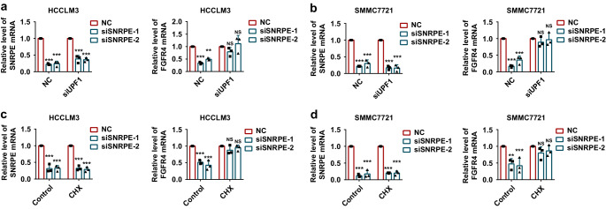
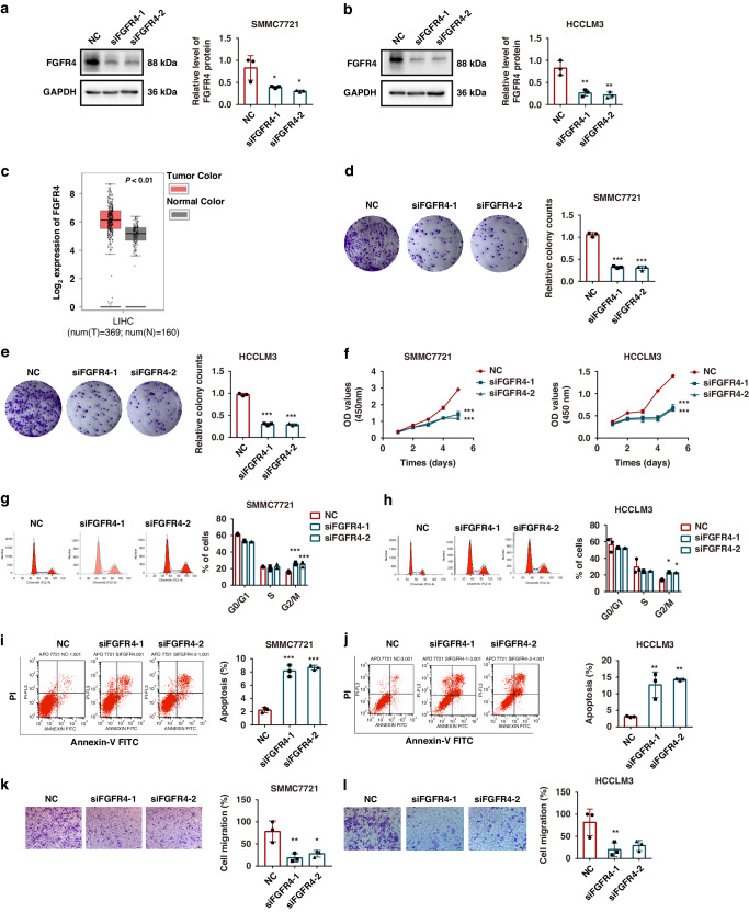
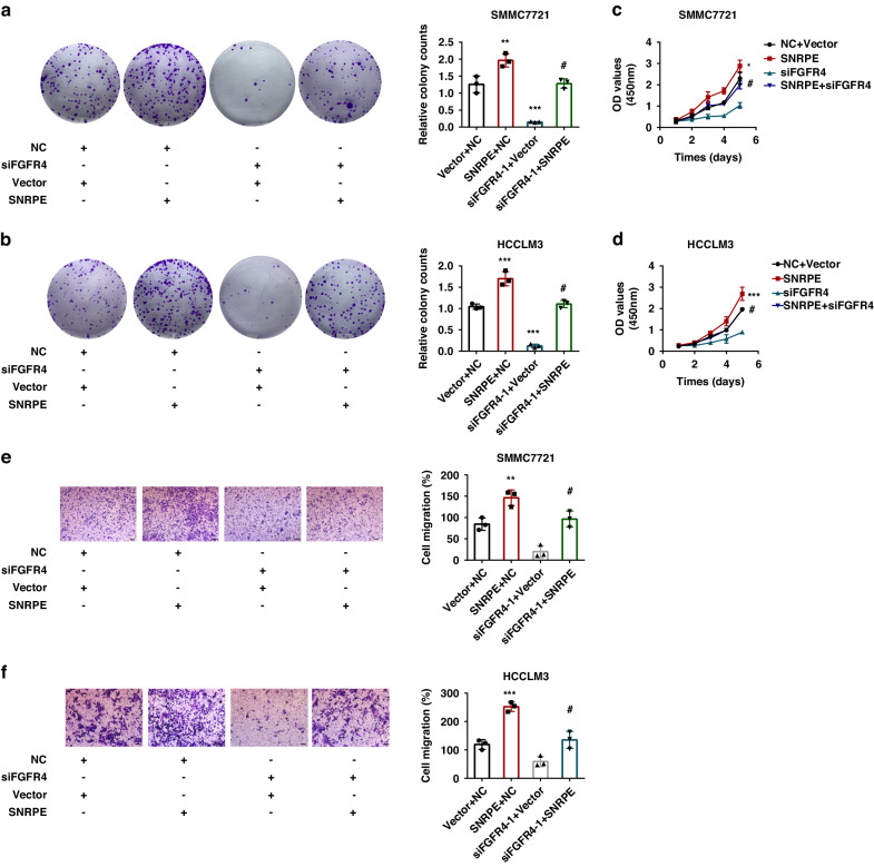
Results: We herein identified SNRPE as a crucial oncofetal splicing factor, significantly associated with the adverse prognosis of HCC. SOX2 was identified as the activator for SNRPE reactivation. Efficient knockdown of SNRPE resulted in the complete cessation of HCC tumorigenesis and progression. Mechanistically, SNRPE knockdown reduced FGFR4 mRNA expression by triggering nonsense-mediated RNA decay. A partial inhibition of SNRPE-induced malignant progression of HCC cells was observed upon FGFR4 knockdown.

Conclusions: Our findings highlight SNRPE as a novel oncofetal splicing factor and shed light on the intricate relationship between oncofetal splicing factors, splicing events, and carcinogenesis. Consequently, SNRPE emerges as a potential therapeutic target for HCC treatment. Model of oncofetal SNRPE promotes HCC tumorigenesis by regulating the AS of FGFR4 pre-mRNA.


Conflict of interest statement

The authors declare no competing interests.

Figures

None
Model of oncofetal SNRPE promotes HCC tumorigenesis by regulating the AS of FGFR4 pre-mRNA.
Fig. 1
Fig. 1. SNRPE, an oncofetal protein, is upregulated by SOX2.
a Candidate oncofetal genes were identified with reference to the GEO and TCGA datasets. b The SNRPE expressions were analyzed in mouse liver development from GSE21224 and GSE13149 datasets. c The SNRPE expressions were analyzed in human liver development from the GSE130473 data. d, e SNRPE mRNA and protein levels were analyzed by PCR and western blotting in mouse fetal liver and adult hepatic tissues. f The SNRPE expressions were analyzed in human HCC tissues (n = 369 independent samples) and normal hepatic tissues (n = 160 independent samples) with reference to the GEPIA database. g The SNRPE expressions were analyzed in normal hepatic tissues and adjacent HCC tumor from GSE121248 and GSE14520 datasets. h SNRPE staining was assessed in human HCC tissues (n = 90 independent samples) and the adjacent normal tissues (n = 85 independent samples) by tissue microarray. Scale bars: 50 μm. i, j The correlation between the SNRPE protein levels and overall survival or recurrence-free survival was analyzed by Kaplan–Meier. k The protein levels of SOX2 were evaluated in SOX2-silenced cells. l The protein levels of SNRPE were detected in SOX2-silenced HCCLM3 cells. m 293T cells were transduced with vector or SOX2-overexpressed plasmid. Simultaneously, the cells were co-transduced with the pGL3-SNRPE (−2000/ + 1) and pRL-TK plasmids. The dual-luciferase activity was determined. The results are expressed as the mean ± SD, n = 6 for (e) and n = 3 for (km). *P < 0.05, **P < 0.01, ***P < 0.001 were determined by Student’s t test.
Fig. 2
Fig. 2. SNRPE promotes HCC carcinogenesis.
a The protein levels of SNRPE were analyzed in SNRPE-overexpressed SMMC7721 or HL7702 cells. b Vector or SNRPE-overexpressed SMMC7721 and HL7702 cells were subjected to a soft agar colony assay. Scale bars: 125 μm, 25 μm. c The cell proliferation of SNRPE-overexpressed SMMC7721 or HL7702 cells was evaluated by colony formation assay. d The cell proliferation of SNRPE-overexpressed SMMC7721 or HL7702 cells was assessed by cell growth curves obtained from the CCK-8 assays. The results are expressed as the mean ± SD, n = 3. *P < 0.05, **P < 0.01, ***P < 0.001 were determined by Student’s t test. eg The effects of SNRPE-overexpressed SMMC7721 and HL7702 cells on subcutaneous tumor growth and tumor volumes were observed every 3 days. The mice were sacrificed 4 weeks after injection. Then, the tumors were excised and weighed (n = 10 for each group). The results are expressed as the mean ± SD. *P < 0.05, **P < 0.01, ***P < 0.001 were determined by Student’s t test. h, i The cell proliferative ability of subcutaneous tumor tissues was evaluated by Ki67 IHC staining. Scale bars: 50 μm, 25 μm.
Fig. 3
Fig. 3. SNRPE is required for tumorigenesis of HCCLM3 cells.
a The protein levels of SNRPE were analyzed in NC or SNRPE-transfected HCCLM3 cells. b NC or SNRPE-transfected HCCLM3 cells were subjected to a soft agar colony assay. Scale bars: 125 μm, 25 μm. c, d The cell proliferation was analyzed by cell growth curve and colony formation assay in NC or SNRPE-transfected HCCLM3 cells. e Cell proliferation was analyzed by EdU immunofluorescence staining in SNRPE-silenced HCCLM3 cells. Scale bars: 25 μm. f The migration of SNRPE-silenced HCCLM3 cells by transwell assays. Scale bars: 50 μm. g Gene Set Enrichment Analyses (GSEA) of the LIHC TCGA dataset. h Cell cycle assays were performed by flow cytometry in control or SNRPE-silenced HCCLM3 cells, followed by quantification of G1 or G2 or S phase cells. i Cell apoptosis of the control and SNRPE-silenced HCCLM3 cells was assessed by flow cytometry, followed by quantification of apoptosis. Data are presented as the mean ± SD, n = 3. *P < 0.05, **P < 0.01, ***P < 0.001 were calculated by one-way ANOVA with Dunnett’s post-test. j, k Effects of SNRPE-silenced HCCLM cells on subcutaneous tumor growth and tumor volumes were analyzed every 3 days. The mice were sacrificed 4 weeks after injection. Then, the tumors were excised and weighed (n = 10 for each group). l Lung metastasis by tail vein xenograft of HCCLM3-shNC cells or HCCLM3-shSNRPE cells (n = 9 for each group). Scale bars: 25 μm. The results are expressed as the mean ± SD. ***P < 0.001 were determined by Student’s t test.
Fig. 4
Fig. 4. SNRPE regulates the alternative splicing of FGFR4 and CREB3L4 mRNA in SMMC7721 and HCCLM3 cells.
a AS events controlled by SNRPE were quantified. SE, skipped exon; MEX, mutually exclusive exon; IR, intron retention; A5SS, alternative 5’ splicing site; A3SS, alternative 3’ splicing site. b KEGG enrichment analysis of SNRPE-regulated gene expression events. c Inclusions of FGFR4-IR and CREB3L4-IR were examined in SNRPE-silenced SMMC7721 or HCCLM3 cells by RT-PCR. d The correlation between SNRPE and FGFR4 levels in human HCC tissues was analyzed from cbioportal. P < 0.001 and r = 0.5374, by Pearson’s correlational analysis. The correlation between SNRPE and CREB3L4 levels in human HCC tissues was analyzed from cbioportal. P < 0.001 and r = 0.5374, by Pearson’s correlational analysis. e The mRNA levels of SNRPE or FGFR4 in SNRPE-silenced SMMC7721 or HCCLM3 cells were evaluated. f The mRNA levels of SNRPE or CREB3L4 in SNRPE-silenced SMMC7721 or HCCLM3 cells were evaluated. g, h The protein levels of FGFR4 in SNRPE-silenced SMMC7721 or HCCLM3 cells were observed by western blotting. Data are presented as the mean ± SD, n = 3. *P < 0.05, **P < 0.01, ***P < 0.001 were calculated by one-way analysis of variance with Dunnett’s post-test.
Fig. 5
Fig. 5. SNRPE regulates FGFR4 expression by activating NMD.
The mRNA levels of SNRPE or FGFR4 were examined in SNRPE-silenced HCCLM3 (a) or SMMC7721 cells (b) transfected with siUPF1. The mRNA levels of SNRPE or FGFR4 were examined in SNRPE-silenced HCCLM3 (c) or SMMC7721 cells (d) treated with CHX. Data are presented as the mean ± SD, n = 3. **P < 0.01, ***P < 0.001 were calculated by two-way analysis of variance with Dunnett’s multiple comparisons test.
Fig. 6
Fig. 6. The effect of FGFR4 on HCC tumorigenesis in HCC cells.
a, b The protein levels of SNRPE were evaluated in NC or FGFR4-transfected SMMC7721 or HCCLM3 cells. c FGFR4 expressions in human HCC tissues (n = 369 independent samples) and normal hepatic tissues (n = 160 independent samples) were analyzed from GEPIA database. d, e The cell proliferation was examined by colony formation assay in NC or FGFR4- transfected SMMC7721 or HCCLM3 cells. f The cell proliferation was examined by cell growth curve in NC or FGFR4- transfected SMMC7721 or HCCLM3 cells. g, h Cell cycle assays were performed via flow cytometry in NC or FGFR4-silenced SMMC7721 or HCCLM3 cells, followed by quantification of G1 or G2 or S phase cells. i, j Cell apoptosis of NC and FGFR4-silenced SMMC7721 or HCCLM3 cells was determined by flow cytometry and quantification of apoptosis. k, l The migration of FGFR4-silenced SMMC7721 or HCCLM3 cells was examined by transwell assay. Scale bars: 50 μm. Data are presented as the mean ± SD, n = 3. *P < 0.05, **P < 0.01, ***P < 0.001 were calculated by one-way analysis of variance with Dunnett’s post-test.
Fig. 7
Fig. 7. FGFR4 knockdown rescues the biological effects of SNRPE-overexpressed HCC cells.
a, b The cell proliferation was examined by colony formation assay in FGFR4-silenced or SNRPE-overexpressed or FGFR4-silenced and SNRPE-overexpressed SMMC7721 or HCCLM3 cells. c, d The cell proliferation was examined by cell growth curve in FGFR4-silenced or overexpressed SNRPE or FGFR4-silenced and SNRPE-overexpressed SMMC7721 or HCCLM3 cells. e, f The migration of FGFR4-silenced or SNRPE-overexpressed or FGFR4-silenced and SNRPE-overexpressed SMMC7721 or HCCLM3 cells was evaluated by transwell assays. Scale bars: 25 μm. Data are presented as the mean ± SD, n = 3. #P<0.05, *P < 0.05, **P < 0.01, ***P < 0.001 were calculated by two-way analysis of variance with Turkey’s post-test.

Similar articles

Cited by

References

    1. Sung H, Ferlay J, Siegel RL, Laversanne M, Soerjomataram I, Jemal A, et al. Global Cancer Statistics 2020: GLOBOCAN estimates of incidence and mortality worldwide for 36 cancers in 185 countries. CA Cancer J Clin. 2021;71:209–49. doi: 10.3322/caac.21660. - DOI - PubMed
    1. Wang H, Lu Z, Zhao X. Tumorigenesis, diagnosis, and therapeutic potential of exosomes in liver cancer. J Hematol Oncol. 2019;12:1–21. doi: 10.1186/s13045-019-0806-6. - DOI - PMC - PubMed
    1. Xu W, Huang H, Yu L, Cao L. Meta-analysis of gene expression profiles indicates genes in spliceosome pathway are up-regulated in hepatocellular carcinoma (HCC) Med Oncol. 2015;32:96. doi: 10.1007/s12032-014-0425-6. - DOI - PubMed
    1. Matera AG, Wang Z. A day in the life of the spliceosome. Nat Rev Mol Cell Biol. 2014;15:108–21. doi: 10.1038/nrm3742. - DOI - PMC - PubMed
    1. Cieśla M, Ngoc PCT, Cordero E, Martinez ÁS, Morsing M, Muthukumar S, et al. Oncogenic translation directs spliceosome dynamics revealing an integral role for SF3A3 in breast cancer. Mol Cell. 2021;81:1453–68.e1412. doi: 10.1016/j.molcel.2021.01.034. - DOI - PubMed

MeSH terms

Substances