Case Reports
. 2024 May 26:25:e943027.
doi: 10.12659/AJCR.943027.

Severe Complications of Uterine Dehiscence Post-Lower Segment Cesarean Section: A Case Report Emphasizing the Importance of Timely Diagnosis and Intervention

Affiliations
Case Reports

Severe Complications of Uterine Dehiscence Post-Lower Segment Cesarean Section: A Case Report Emphasizing the Importance of Timely Diagnosis and Intervention

Saifon Chawanpaiboon et al. Am J Case Rep. .

Abstract

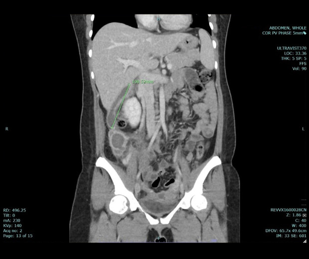
BACKGROUND Uterine dehiscence, an infrequent event often mistaken for uterine rupture, is rarely linked to post-cesarean section procedures and can result in severe complications, notably puerperal sepsis. In this report, we present a case that exemplifies the onset of puerperal sepsis and the emergence of intra-abdominal abscesses attributed to uterine dehiscence following a lower segment cesarean section (LSCS). CASE REPORT Our patient, a 28-year-old woman in her third pregnancy, underwent LSCS 1 week earlier. Subsequently, she returned to the hospital with lower abdominal pains, fever, and malodorous vaginal discharge. Computed tomography (CT) scan of whole abdomen verified uterine dehiscence and pus collection at the subhepatic region and right paracolic gutter. After referral to a specialized hospital, laboratory findings indicated an elevated white blood cell count and alkaline phosphatase levels, and coagulation abnormalities. She underwent an exploratory laparotomy, which unveiled uterine dehiscence, abscesses, and adhesions, necessitating a total abdominal hysterectomy and abdominal toileting. Pus culture analysis identified the presence of E. coli, which was susceptible to ampicillin/sulbactam. Complications were encountered after surgery, including wound dehiscence and pus re-accumulation. Successful management involved vacuum dressings and percutaneous drainage. Eventually, her condition improved and she was discharged, without additional complications. CONCLUSIONS This report underscores the importance of considering cesarean scar dehiscence as a diagnosis in women with previous cesarean deliveries who present during subsequent pregnancies with symptoms such as abdominal pain or abdominal sepsis. Diagnostic tools, such as CT, play pivotal roles, and the timely performance of an exploratory laparotomy is paramount when suspicion arises.


Conflict of interest statement

Conflict of interest: None declared

Figures

Figure 1.
Figure 1.
Previous computed tomography scan of the patient’s entire abdominal region (coronal view). Identification of fluid accumulation with concomitant air bubble formation from the lower uterine segment, raising concerns about potential uterine dehiscence at her prior cesarean section scar site. Remarkable abscess formation, characterized by dimensions of 6.5×1.4×3.6 cm and 2.1×3.4×2.3 cm, noted in the subhepatic region and the right paracolic gutter, measuring 4.6×2.9×15.2 cm and 4.7×4.5×5.6 cm, respectively.
Figure 2.
Figure 2.
Previous computed tomography scan of the patient’s entire abdominal region (sagittal view). Identification of fluid accumulation with concomitant air bubble formation from the lower uterine segment, raising concerns of potential uterine dehiscence at her prior cesarean section scar site. Remarkable abscess formation, characterized by dimensions of 6.5×1.4×3.6 cm and 2.1×3.4×2.3 cm, noted in the subhepatic region and the right paracolic gutter, measuring 4.6×2.9×15.2 cm and 4.7×4.5×5.6 cm, respectively.
Figure 3.
Figure 3.
Intraoperative image depicts a total lower uterine dehiscence spanning 10 cm. This dehiscence is characterized by fragility, indicating weakness in the tissue structure, and the presence of necrotic tissue, suggesting tissue death, both observed at the lower uterine segment and surgical margins.
Figure 4.
Figure 4.
The specimen presented is the body of the uterus with the cervix, specifically showing the posterior surface. It is notable that the uterus is separated into 2 pieces due to the presence of fragile, necrotic tissue at the lower uterine segment. This separation is likely a result of the weakened tissue integrity caused by the necrotic tissue.
Figure 5.
Figure 5.
The specimen presented shows the separation of the uterus into 2 parts, specifically caused by necrotic tissue located at the lower uterine segment. This separation into 2 parts is a consequence of the weakened tissue integrity resulting from the presence of necrotic tissue, which can lead to structural instability and potentially serious complications.
Figure 6.
Figure 6.
After the removal of the Jackson drain, a disruption or separation of the abdominal wound was observed.
Figure 7.
Figure 7.
Postoperative computed tomography scan of the patient’s entire abdominal region (coronal view). A follow-up computed tomography scan revealed the continued presence of pus collection in the subhepatic region and the right paracolic gutter, although it appeared to be smaller in size than the previous scan.
Figure 8.
Figure 8.
The plastic surgeon recommended the use of vacuum dressings with continuous drainage to manage the disrupted wound.
Figure 9.
Figure 9.
An interventional radiologist was engaged to perform percutaneous drainage under ultrasound guidance.
Figure 10.
Figure 10.
After 7 days of vacuum dressing application, significant improvement was noted in the appearance of the abdominal wound. The wound exhibited notable progress, appearing significantly better than its initial state.

References

    1. Rahman M, Khan N, Rahman A, et al. Long-term effects of caesarean delivery on health and behavioural outcomes of the mother and child in Bangladesh. J Health Popul Nutr. 2022;41(1):45. - PMC - PubMed
    1. World Health Orginization Caesarean section rates continue to rise, amid growing inequalities in access. 2021. Available from: https://www.who.int/news/item/16-06-2021-caesarean-section-rates-continu....
    1. Diaz SD, Jones JE, Seryakov M, Mann WJ. Uterine rupture and dehiscence: Ten-year review and case-control study. South Med J. 2002;95(4):431–35. - PubMed
    1. Wagner MS, Bédard MJ. Postpartum uterine wound dehiscence: A case report. J Obstet Gynaecol Can. 2006;28(8):713–15. - PubMed
    1. Royo P, Manero MG, Olartecoechea B, Alcázar JL. Two-dimensional power Doppler-three-dimensional ultrasound imaging of a cesarean section dehiscence with utero-peritoneal fistula: A case report. J Med Case Rep. 2009;3:42. - PMC - PubMed

Publication types