. 2024 May 26;14(1):12016.
doi: 10.1038/s41598-024-62689-6.

Effect of hypercholesterolemia on circulating and cardiomyocyte-derived extracellular vesicles

Affiliations

Effect of hypercholesterolemia on circulating and cardiomyocyte-derived extracellular vesicles

Csenger Kovácsházi et al. Sci Rep. .

Abstract

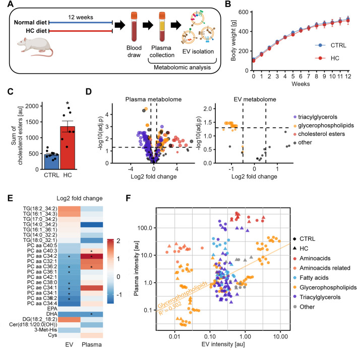
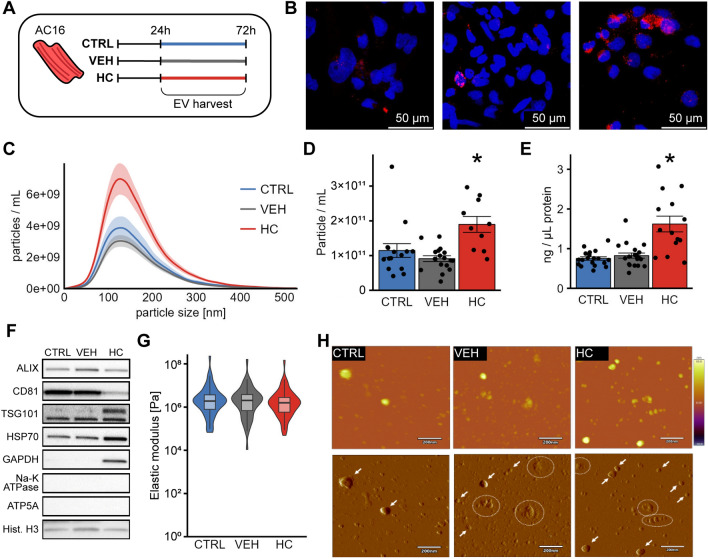
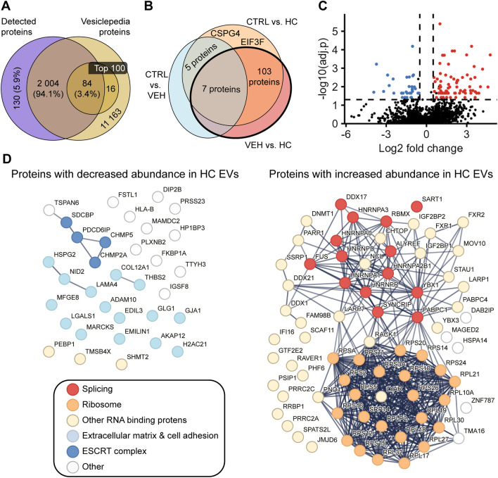
Hypercholesterolemia (HC) induces, propagates and exacerbates cardiovascular diseases via various mechanisms that are yet not properly understood. Extracellular vesicles (EVs) are involved in the pathomechanism of these diseases. To understand how circulating or cardiac-derived EVs could affect myocardial functions, we analyzed the metabolomic profile of circulating EVs, and we performed an in-depth analysis of cardiomyocyte (CM)-derived EVs in HC. Circulating EVs were isolated with Vezics technology from male Wistar rats fed with high-cholesterol or control chow. AC16 human CMs were treated with Remembrane HC supplement and EVs were isolated from cell culture supernatant. The biophysical properties and the protein composition of CM EVs were analyzed. THP1-ASC-GFP cells were treated with CM EVs, and monocyte activation was measured. HC diet reduced the amount of certain phosphatidylcholines in circulating EVs, independently of their plasma level. HC treatment significantly increased EV secretion of CMs and greatly modified CM EV proteome, enriching several proteins involved in tissue remodeling. Regardless of the treatment, CM EVs did not induce the activation of THP1 monocytes. In conclusion, HC strongly affects the metabolome of circulating EVs and dysregulates CM EVs, which might contribute to HC-induced cardiac derangements.

Keywords: Dyslipidemia; Exosome; Inflammation; Metabolomics; Obesity; Proteomics.


Conflict of interest statement

PF is the founder and CEO, and ZG is the Translational Program Director of Pharmahungary Group, a group of R&D companies. EIB is member of the Advisory Board of Sphere Gene Therapeutics Inc. (Boston, MA, USA) and the Scientific Advisory Board of ReNeuron (UK). All other authors declare no competing interests.

Figures

Figure 1
Figure 1
Metabolomic analysis of circulating EVs in HC-fed rats. (nctrl = 11, nhc = 7 for all experiments) (A) Schematic representation of the experimental plans. Male Wistar rats were kept on normal or HC diet for 12 weeks, then blood was collected and EVs were isolated from platelet-free plasma. Plasma and EV metabolome were analyzed. (B) Body weight of the animals. Diet did not affect body weight. (C) HC diet increased the amount of cholesterol esters in the blood. (D) Volcano plot of metabolome analysis on plasma (left) and EVs (right). In plasma, the intensity of numerous triacylglycerols was significantly reduced, meanwhile glycerophospholipids and cholesterol esters were enriched by HC. In EVs, the intensity of most glycerophospholipids detected was decreased by HC. (E) Heatmap for the metabolites detected both in EVs and in plasma. Multiple metabolites show opposite changes between EVs and plasma. (F) Linear correlation of multiple metabolites in EVs vs. plasma. Of the five metabolite groups analyzed, only glycerophospholipids showed a moderate correlation between plasma and EV intensities (see regression line, R2 = 0.502, p < 0.001). *p < 0.05 Student’s t-test CTRL vs HC; For metabolomics analysis (panel E) Benjamini–Hochberg false discovery p-adjustment was applied.
Figure 2
Figure 2
Analysis of EVs secreted by AC16 CMs. (A) Schematic representation of the experimental plans. AC16 human CMs were seeded for 24 h, and then the medium was replaced with serum-free medium with or without vehicle or HC supplement for another 48 h, then EVs were isolated from the cell culture supernatant. (B) Representative images of oil-red-o measurements on AC16 cells. Blue: DAPI (nuclei), red: Oil-red-O (lipid). Results show increased amounts of lipids upon HC treatment. (C) Size distribution of isolated EVs measured by NTA. Vesicles with diameters of 50–300 nm were detected in every experimental group. (nctrl = 15, nveh = 17, nhc = 10) (D) Particle number of EV isolates measured by NTA. HC increased the particle number significantly. (nctrl = 15, nveh = 17, nhc = 10) (E) Absolute protein concentration of the isolates measured with 280 nm light absorbance. HC increased the protein concentration significantly. (nctrl = 19, nveh = 20, nhc = 14) (F) Analysis of EV markers and potential contaminants using Western blot. Whole blots are presented in Supplementary Figure S3. (G) Elastic modulus of the isolates measured by AFM. No significant difference was observed. (nctrl = 58, nveh = 49, nhc = 54, out of at least three independent experiments) (H) Representative images of non-contact mode AFM measurements. First row: height-contrast, second row: amplitude-contrast images in the same view field. Arrows: EVs, Circles: Non-EV-like structures, with height < 10 nm. *p < 0.05 HC vs CTRL and VEH in ANOVA with Tukey’s post-hoc test.
Figure 3
Figure 3
Analysis of immune cell activation of AC16 EVs using THP1-ASC-GFP. (A) Schematic representation of the experimental setup. EVs were isolated from AC16 cells and THP1-ASC-GFP cells were treated with the isolates, then flow cytometry measurement was applied to measure GFP expression and protein expression was analyzed with qPCR. (B) GFP expression measured by flow cytometry. LPS significantly increased the percentage of activated cells. EV treatment did not affect GFP expression, regardless of the dose or the experimental group (n = 6, all groups). (C) Gene expression analysis of IL-1β, TNF-α and IL-10 normalized to HPRT. No differences were observed (n = 2–3) *p < 0.05 vs PBS; ANOVA with Tukey’s post-hoc test.
Figure 4
Figure 4
Proteomic analysis of AC16 EVs. (nctrl = 10, nveh = 10, nhc = 4, in all experiments) (A) Comparison of the proteins detected with the Vesiclepedia database. A total of 2137 proteins were identified of which 2088 were described in the database and 84 proteins were found among the 100 most frequently described EV proteins. (B) Venn diagram for the representation of the statistically significant differences between the experimental groups, using ANOVA followed by Turkey’s post-hoc test. (C) Volcano plot shows significant differences between the VEH and HC groups. (D) Interaction network of proteins with significantly different abundance between the VEH and HC groups. LEFT: Proteins with decreased abundance, RIGHT: Proteins with increased abundance.

References

    1. Murray CJL. The global burden of disease study at 30 years. Nat. Med. 2022;28:2019–2026. doi: 10.1038/s41591-022-01990-1. - DOI - PubMed
    1. Théry C, et al. Minimal information for studies of extracellular vesicles 2018 (MISEV2018): A position statement of the international society for extracellular vesicles and update of the MISEV2014 guidelines. J. Extracell. Vesicles. 2018;7:1535750. doi: 10.1080/20013078.2018.1535750. - DOI - PMC - PubMed
    1. Sluijter JPG, et al. Extracellular vesicles in diagnostics and therapy of the ischaemic heart: Position paper from the working group on cellular biology of the heart of the European society of cardiology. Cardiovasc. Res. 2018;114:19–34. doi: 10.1093/cvr/cvx211. - DOI - PMC - PubMed
    1. Akbar N, Azzimato V, Choudhury RP, Aouadi M. Extracellular vesicles in metabolic disease. Diabetologia. 2019;62:2179. doi: 10.1007/s00125-019-05014-5. - DOI - PMC - PubMed
    1. Ferdinandy P, Hausenloy DJ, Heusch G, Baxter GF, Schulz R. Interaction of risk factors, comorbidities, and comedications with ischemia/reperfusion injury and cardioprotection by preconditioning, postconditioning, and remote conditioning. Pharmacol. Rev. 2014;66:1142–1174. doi: 10.1124/pr.113.008300. - DOI - PubMed