. 2024 May 27;57(1):32.
doi: 10.1186/s40659-024-00510-4.

The hepatoprotective effect of 4-phenyltetrahydroquinolines on carbon tetrachloride induced hepatotoxicity in rats through autophagy inhibition

Affiliations

The hepatoprotective effect of 4-phenyltetrahydroquinolines on carbon tetrachloride induced hepatotoxicity in rats through autophagy inhibition

Mohamed Hussein Abdelgalil et al. Biol Res. .

Abstract

Background: The liver serves as a metabolic hub within the human body, playing a crucial role in various essential functions, such as detoxification, nutrient metabolism, and hormone regulation. Therefore, protecting the liver against endogenous and exogenous insults has become a primary focus in medical research. Consequently, the potential hepatoprotective properties of multiple 4-phenyltetrahydroquinolines inspired us to thoroughly study the influence of four specially designed and synthesized derivatives on carbon tetrachloride (CCl4)-induced liver injury in rats.

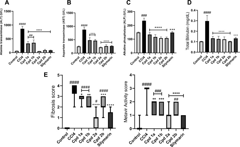
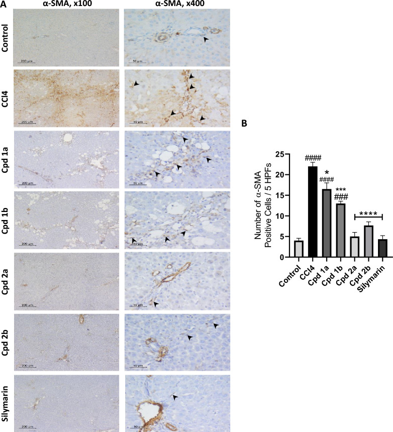
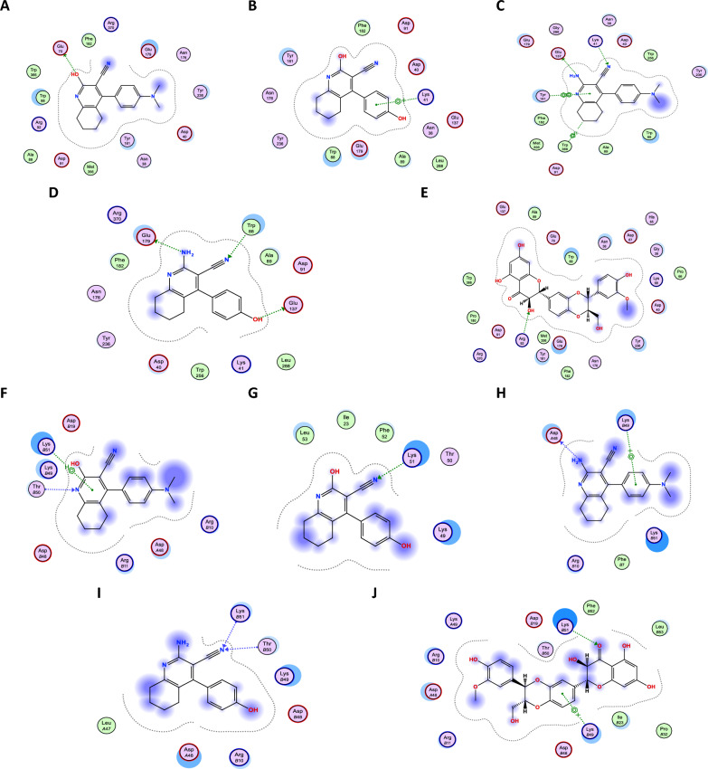
Methods and results: Seventy-seven Wistar albino male rats weighing 140 ± 18 g were divided into eleven groups to investigate both the toxicity profile and the hepatoprotective potential of 4-phenyltetrahydroquinolines. An in-vivo hepatotoxicity model was conducted using CCl4 (1 ml/kg body weight, a 1:1 v/v mixture with corn oil, i.p.) every 72 h for 14 days. The concurrent treatment of rats with our newly synthesized compounds (each at a dose of 25 mg/kg body weight, suspended in 0.5% CMC, p.o.) every 24 h effectively lowered transaminases, preserved liver tissue integrity, and mitigated oxidative stress and inflammation. Moreover, the histopathological examination of liver tissues revealed a significant reduction in liver fibrosis, which was further supported by the immunohistochemical analysis of α-SMA. Additionally, the expression of the apoptotic genes BAX and BCL2 was monitored using real-time PCR, which showed a significant decrease in liver apoptosis. Further investigations unveiled the ability of the compounds to significantly decrease the expression of autophagy-related proteins, Beclin-1 and LC3B, consequently inhibiting autophagy. Finally, our computer-assisted simulation dockingonfirmed the obtained experimental activities.

Conclusion: Our findings suggest that derivatives of 4-phenyltetrahydroquinoline demonstrate hepatoprotective properties in CCl4-induced liver damage and fibrosis in rats. The potential mechanism of action may be due to the inhibition of autophagy in liver cells.

Keywords: 4-phenyltetrahydroquinoline; Autophagy; CCL4-induced hepatotoxicity; CYP2E1; HepG2; Hepatoprotective; Liver injury; Tacrine derivatives.


Conflict of interest statement

The authors declare no competing interests.

Figures

Fig. 1
Fig. 1
Rational for the synthesis of compounds 1a, 1b, 2a, and 2b
Fig. 2
Fig. 2
The effect of Cpds (1a, 1b, 2a, and 2b) on HepG2 cells. IC50 values in μM are represented as mean ± SEM. *p < 0.05, **p < 0.01, ***p < 0.001 compared to tacrine
Fig. 3
Fig. 3
The effect of Cpds (1a, 1b, 2a, and 2b) on the Safety Profile. A Liver function tests (ALT and AST). B Lipid profile (total cholesterol and triglycerides). C Kidney function tests (serum urea and creatinine). Values are represented as the mean ± SEM, n = 7. *p < 0.05, **p < 0.01
Fig. 4
Fig. 4
Histopathological assessment of the safety of Cpds (1a, 1b, 2a, and 2b) on liver tissues. H&E stained liver sections (n = 5) show normal liver architecture and normal hepatocytes. Mild portal or lobular inflammation is seen occasionally in Cpd 1b and 2b (arrow). Masson trichrome-stained sections (n = 5) show no fibrosis (F0) in low power. High power view of one portal tract shows scanty blue-stained fibrous tissue around portal vessels. Scale bar for low power (× 100) is 200 μM and for high power (× 400) is 50 μM
Fig. 5
Fig. 5
The Effect of Cpds (1a, 1b, 2a, and 2b) on Liver Biomarkers, Degree of Fibrosis and Metavir Activity Scores. A Alanine transaminase. B Aspartate transaminase. C Alkaline phosphatase. D total bilirubin. Values are represented as the mean ± SEM, n = 7. E The degree of fibrosis and Metavir activity scores in liver tissues using the Metavir scoring system. Results are represented as medians (minimum to maximum), box and whiskers. *p < 0.05, **p < 0.01, ***p < 0.001, ****p < 0.0001 compared to the hepatotoxicity group. #p < 0.05, ##p < 0.01, ###p < 0.001, ####p < 0.0001 compared to the control group
Fig. 6
Fig. 6
Histopathological assessment of Liver Fibrosis and Metavir activity. Liver sections stained with H&E to assess liver activity, and sections stained with Masson trichrome to assess the degree of fibrosis, n = 7. H&E high power (× 400) shows macrovesicular steatosis (arrows) and apoptotic cells (arrow heads), as well as foci of inflammation (stars). In Masson trichrome stained sections, P portal tract, C central vein, arrow fibrous bridging. Scale bar for low power (× 100) is 200 μM and for high power (× 200) is 100 μM and (× 400) is 50 μM
Fig. 7
Fig. 7
The Effect of Cpds (1a, 1b, 2a and 2b) on oxidative stress, inflammation, fibrosis, and apoptosis. Oxidative stress markers, A MDA, and B GSH levels were determined spectrophotometrically. The levels of pro-inflammatory, and pro-fibrotic cytokines, C TNF-α and D TGF-β were quantified using ELISA technique. E The level of expression of both the pro-apoptotic and the anti-apoptotic genes, BAX and BCL2, respectively, was determined using qPCR and the ratio of BAX/BCL2 was calculated. Values are represented as mean ± SEM, n = 7. *p < 0.05, ***p < 0.001, ****p < 0.0001 compared to hepatotoxicity group. ###p < 0.001, ####p < 0.0001 compared to control group
Fig. 8
Fig. 8
Immunohistochemical assessment of α-SMA in liver tissues. A Assessment of α-SMA positive cells in α-SMA immune stained liver section, n = 3. Blood vessels in portal tracts are stained positive as an internal positive control. Arrows are pointing at the positively stained HSCs/myofibroblasts. They are seen as long flat cells in between hepatocytes and within portal tracts. Scale bar for low power (× 100) is 200 μM and for high power (× 400) is 50 μM. B Number of α-SMA positive cells / 5 HPFs. Values are represented as mean ± SEM. *p < 0.05, ***p < 0.001, ****p < 0.0001 compared to the hepatotoxicity group. ###p < 0.001, ####p < 0.0001 compared to the control group
Fig. 9
Fig. 9
The Effect of Cpds (1a, 1b, 2a and 2b) on CYP2E1 and Autophagy. A The levels of CYP2E1 enzyme expression. B Autophagy-related proteins, Beclin-1 and LC3B. Protein expression is measured using ELISA technique. Values are represented as mean ± SEM, n = 7. ****p < 0.0001 compared to hepatotoxicity group. #p < 0.05, ##p < 0.01, ###p < 0.001, ####p < 0.0001 compared to control group
Fig. 10
Fig. 10
2D Binding mode of compounds 1a, 1b, 2a, 2b and Silymarin in the binding site of Beclin-1 and LC3B receptors. A Compound 1a binding to Beclin-1 receptor. B Compound 1b binding to Beclin-1 receptor. C Compound 2a binding to Beclin-1 receptor. D Compound 2b binding to Beclin-1 receptor. E Silymarin binding to Beclin-1 receptor. F Compound 1a binding to LC3B receptor. G Compound 1b binding to LC3B receptor. H Compound 2a binding to LC3B receptor. I Compound 2b binding to LC3B receptor. J Silymarin binding to LC3B receptor
Fig. 11
Fig. 11
2D Binding mode of compounds 1a, 1b, 2a, 2b and Silymarin in the binding site of CYP2E1 receptor. A Compound 1a. B Compound 1b. C Compound 2a. D Compound 2b. E Silymarin

Similar articles

References

    1. Trefts E, Gannon M, Wasserman DH. The liver. Curr Biol. 2017;27(21):R1147–R1151. doi: 10.1016/j.cub.2017.09.019. - DOI - PMC - PubMed
    1. Moreira PR, Maioli MA, Medeiros HCD, Guelfi M, Pereira FTV, Mingatto FE. Protective effect of bixin on carbon tetrachloride-induced hepatotoxicity in rats. Biol Res. 2014;47(1):49. doi: 10.1186/0717-6287-47-49. - DOI - PMC - PubMed
    1. Zhao M, Ma J, Li M, Zhang Y, Jiang B, Zhao X, et al. Cytochrome P450 enzymes and drug metabolism in humans. Int J Mol Sci. 2021 doi: 10.3390/ijms222312808. - DOI - PMC - PubMed
    1. Harjumäki R, Pridgeon CS, Ingelman-Sundberg M. CYP2E1 in alcoholic and non-alcoholic liver injury. Roles of ROS, reactive intermediates and lipid overload. Int J Mol Sci. 2021 doi: 10.3390/ijms22158221. - DOI - PMC - PubMed
    1. Massart J, Begriche K, Hartman JH, Fromenty B. Role of mitochondrial cytochrome P450 2E1 in healthy and diseased liver. Cells. 2022 doi: 10.3390/cells11020288. - DOI - PMC - PubMed