. 2024 Jul 8;227(3):iyae071.
doi: 10.1093/genetics/iyae071.

FLN-2 functions in parallel to linker of nucleoskeleton and cytoskeleton complexes and CDC-42/actin pathways during P-cell nuclear migration through constricted spaces in Caenorhabditis elegans

Affiliations

FLN-2 functions in parallel to linker of nucleoskeleton and cytoskeleton complexes and CDC-42/actin pathways during P-cell nuclear migration through constricted spaces in Caenorhabditis elegans

Linda Ma et al. Genetics. .

Abstract

Nuclear migration through narrow constrictions is important for development, metastasis, and proinflammatory responses. Studies performed in tissue culture cells have implicated linker of nucleoskeleton and cytoskeleton (LINC) complexes, microtubule motors, the actin cytoskeleton, and nuclear envelope repair machinery as important mediators of nuclear movements through constricted spaces. However, little is understood about how these mechanisms operate to move nuclei in vivo. In Caenorhabditis elegans larvae, six pairs of hypodermal P cells migrate from lateral to ventral positions through a constricted space between the body wall muscles and the cuticle. P-cell nuclear migration is mediated in part by LINC complexes using a microtubule-based pathway and by an independent CDC-42/actin-based pathway. However, when both LINC complex and actin-based pathways are knocked out, many nuclei still migrate, suggesting the existence of additional pathways. Here, we show that FLN-2 functions in a third pathway to mediate P-cell nuclear migration. The predicted N-terminal actin-binding domain in FLN-2 that is found in canonical filamins is dispensable for FLN-2 function; this and structural predictions suggest that FLN-2 does not function as a filamin. The immunoglobulin-like repeats 4-8 of FLN-2 were necessary for P-cell nuclear migration. Furthermore, in the absence of the LINC complex component unc-84, fln-2 mutants had an increase in P-cell nuclear rupture. We conclude that FLN-2 functions to maintain the integrity of the nuclear envelope in parallel with the LINC complex and CDC-42/actin-based pathways to move P-cell nuclei through constricted spaces.

Keywords: Cdc42; LINC complexes; enhancers; filamin; hypodermal development; nuclear migration.


Conflict of interest statement

Conflicts of interest The author(s) declare no conflict of interest.

Figures
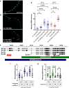
Fig. 1.
Fig. 1.
fln-2 mutants enhance the P-cell nuclear migration defect observed in unc-84 worms. a–c) Representative epifluorescence images of unc-47::gfp-marked GABA neurons in wild-type, unc-84(n369), or unc-84(n369); fln-2(tm4687) animals. d) The numbers of missing GABA neurons are shown from the following genotypes: unc-84(n369) (black), fln-2(tm4687) (magenta), unc-84(n369) fln-2(tm4687) (red), and unc-84(n369) fln-2(RNAi) (pink). **** signifies a P-value < 0.0001 when compared with unc-84(n369) alone. e) Schematic of four of the more than 25 predicted isoforms of fln-2. The purple diamond indicates the ot611 point mutant producing an early stop codon. The pink box indicates the X-4P01 clone used for RNAi. The magenta box shows the location of the tm4687 deletion allele. The fosmids used in these experiments are marked below the isoforms. The red box marks sequences encoding a predicted actin-binding domain of three calponin homology repeats that is only present in the longer isoforms. f–g) Missing GABA neuron counts for the unc-84(n369) fln-2(tm4687) mutants with and without the indicated fosmids. Rescue experiments were performed on animals grown at 15, 20, or 25°C as indicated below the graphs. + indicates the presence of the extrachromosomal array carrying the fosmid(s) and an RFP, while- indicates there was no fosmid(s) present. *, **, ***, and **** indicate a P-value of <0.02, <0.003, 0.0007, and <0.0001, respectively; error bars are 95% confidence intervals.
Fig. 2.
Fig. 2.
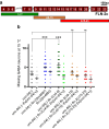
Deletion of Ig-like repeats 4–9 enhances the unc-84 P-cell nuclear migration defect. a) Schematic of the FLN-2c isoform, which contains 20 predicted Ig-like repeats (maroon boxes). Ig-like repeats 1–3 are only found in the longer isoforms of FLMN-2 with interspersed disordered regions indicated by the zigzag lines. The region between repeats 6 and 7 is predicted to be disordered. The deleted portions of FLN-2 in three strains made by CRISPR/Cas9 editing are indicated below the schematic. The scale bar indicates 100 amino acids. b) Missing GABA neurons at 15°C were scored as a proxy for failed P-cell nuclear migrations. See Table 1 for details on the genotypes of the strains used here. Means with 95% confidence intervals are shown. *** indicates significant differences with a P-value of <0.001 when compared with unc-84 null animals, and ns stands for no significant difference from unc-84 null animals. A t-test was performed to generate P-values.
Fig. 3.
Fig. 3.
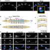
Actin networks during P-cell nuclear migration. a) Representative Airyscan super-resolution images of a wild-type L1 larvae during P-cell nuclear migration. P-cell nuclei are marked by nls::tdTomato (magenta), and actin in P cells is tagged with vab-10::mVenus (cyan). The gold box indicates a P-cell mid-nuclear migration that is enlarged in the inset below. The orange arrow points out actin perpendicular to the direction of nuclear migration in the lateral compartment, and the white arrow indicates actin parallel to the direction of nuclear migration in the constriction. A ventral view is shown, and the ventral cord is marked by the dashed line. anterior is left; scale bar is 10 mm. b and c) A ventral view (b) and lateral view (c) of P cells during the narrowing (b and c) and migration (b′ and c′) stages. P cells are in cyan; nuclei are in magenta. The dashed line indicates the ventral cord. The pharynx indicates the anterior end of the worm, and body wall muscles are represented by the transparent gray bar. d) Actin (vab10::mVenus; cyan) and P-cell nuclei (nls::tdTomato; magenta) in wild-type, unc-84(n369), fln-2(tm4687), or fln-2(tm4687) unc-84(n369) double-mutant animals. Maximum projections of z stacks are shown. The scale bar is 10 mm.
Fig. 4.
Fig. 4.
Localization of FLN-2::spGFP in larval P cells. a) Representative Airyscan super-resolution image of fln-2c::GFP expressed in a C. elegans L1 larva. A lateral view is shown; ventral is toward the lower right. Scale bar is 10 mm. b) Graph depicting the number of missing GABA neurons in unc-84(n369), unc-84(n369) + Ex[unc-84(+)], fln-2c::spGFP unc-84(n369), and fln-2c::spGFP unc-84(n369) + Ex[unc-84(+)] worms. Error bars are 95% CI, and nonsignificant P-values > 0.05.
Fig. 5.
Fig. 5.
Alphafold predictions of the structure of FLN-1 and FLN-2. The highest confidence of five FLN-1a (top) and FLN-2d (bottom) structures predicted by ColabFold version 1.5.2 is shown. On the right, the pLDDT scores (a per-residue confidence metric) for five different predictions are shown. The protein diagrams are colored by sequence order from blue (N terminus) to red (C terminus).
Fig. 6.
Fig. 6.
P-cell nuclei in fln-2  unc-84 mutant animals often rupture during migration. a) Representative Airyscan super-resolution images of P cells late in L1 after or during nuclear migration into the ventral cord in wild-type, unc-84(n369), fln-2(tm4687), and fln-2(tm4687) unc-84(n369) animals. Actin is shown to mark the shape of P cells. Arrows point to suspected nuclear rupture events where the nls::tdTomato marker has leaked into the cytoplasm of the P cell. Maximum projections of z stacks are shown. The scale bar is 10 mm. b) The number of P cells with cytoplasmic nls::tdTomato per worm in late L1. Nuclear rupture was assayed at about 20°C, room temperature for the microscope room. Means and 95% confidence intervals are shown. **** denotes a P-value of <0.00001.
Fig. 7.
Fig. 7.
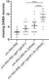
fln-2 has synthetic interactions with unc-84 and cgef-1. Graph depicting the number of missing GABA neurons in the designated genotypes. The mean is shown with 95% confidence interval error bars. Data were collected at 15°C. n is at least 15. *** denotes P < 0.001 from a paired t-test.

Update of

Similar articles

Cited by

References

    1. Altun ZF, Hall DH. 2024. Handbook of C. elegans Anatomy. In WormAtlas.
    1. Arribere JA, Bell RT, Fu BXH, Artiles KL, Hartman PS, Fire AZ. 2014. Efficient marker-free recovery of custom genetic modifications with CRISPR/Cas9 in Caenorhabditis elegans. Genetics. 198(3):837–846. doi:10.1534/genetics.114.169730. - DOI - PMC - PubMed
    1. Bone CR, Chang Y-T, Cain NE, Murphy SP, Starr DA. 2016. Nuclei migrate through constricted spaces using microtubule motors and actin networks in C. elegans hypodermal cells. Development. 143(22):4193–4202. doi:10.1242/dev.141192. - DOI - PMC - PubMed
    1. Bone CR, Starr DA. 2016. Nuclear migration events throughout development. J Cell Sci. 129(10):1951–1961. doi:10.1242/jcs.179788. - DOI - PMC - PubMed
    1. Bone CR, Tapley EC, Gorjanacz M, Starr DA. 2014. The Caenorhabditis elegans SUN protein UNC-84 interacts with lamin to transfer forces from the cytoplasm to the nucleoskeleton during nuclear migration. Mol Biol Cell. 25(18):2853–2865. doi:10.1091/mbc.e14-05-0971. - DOI - PMC - PubMed

MeSH terms