

Learning of the same task subserved by substantially different mechanisms between patients with body dysmorphic disorder and healthy individuals
- PMID: 38798001
- PMCID: PMC11128689
- DOI: 10.1093/cercor/bhae215
Learning of the same task subserved by substantially different mechanisms between patients with body dysmorphic disorder and healthy individuals
Abstract
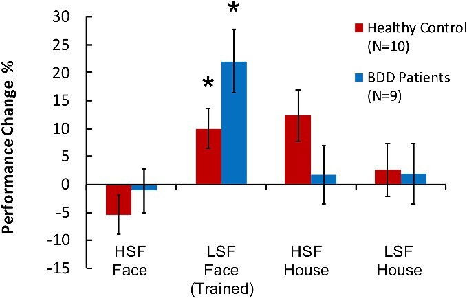
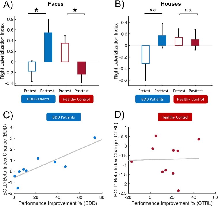
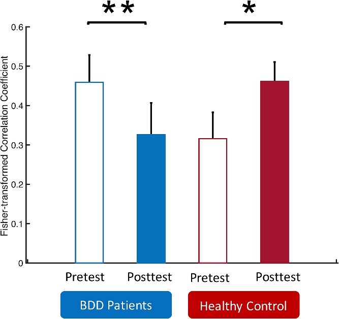
It has remained unclear whether individuals with psychiatric disorders involving altered visual processing employ similar neuronal mechanisms during perceptual learning of a visual task. We investigated this question by training patients with body dysmorphic disorder, a psychiatric disorder characterized by distressing or impairing preoccupation with nonexistent or slight defects in one's physical appearance, and healthy controls on a visual detection task for human faces with low spatial frequency components. Brain activation during task performance was measured with functional magnetic resonance imaging before the beginning and after the end of behavioral training. Both groups of participants improved performance on the trained task to a similar extent. However, neuronal changes in the fusiform face area were substantially different between groups such that activation for low spatial frequency faces in the right fusiform face area increased after training in body dysmorphic disorder patients but decreased in controls. Moreover, functional connectivity between left and right fusiform face area decreased after training in patients but increased in controls. Our results indicate that neuronal mechanisms involved in perceptual learning of a face detection task differ fundamentally between body dysmorphic disorder patients and controls. Such different neuronal mechanisms in body dysmorphic disorder patients might reflect the brain's adaptations to altered functions imposed by the psychiatric disorder.
Keywords: body dysmorphic disorder; fusiform face area; plasticity; visual perceptual learning.
© The Author(s) 2024. Published by Oxford University Press. All rights reserved. For permissions, please e-mail: journals.permissions@oup.com.
Figures
Update of
-
Learning of the same task subserved by substantially different mechanisms between patients with Body Dysmorphic Disorder and healthy individuals.bioRxiv [Preprint]. 2023 Dec 20:2023.12.19.571882. doi: 10.1101/2023.12.19.571882. bioRxiv. 2023. Update in: Cereb Cortex. 2024 May 2;34(5):bhae215. doi: 10.1093/cercor/bhae215. PMID: 38187719 Free PMC article. Updated. Preprint.
Similar articles
-
Learning of the same task subserved by substantially different mechanisms between patients with Body Dysmorphic Disorder and healthy individuals.bioRxiv [Preprint]. 2023 Dec 20:2023.12.19.571882. doi: 10.1101/2023.12.19.571882. bioRxiv. 2023. Update in: Cereb Cortex. 2024 May 2;34(5):bhae215. doi: 10.1093/cercor/bhae215. PMID: 38187719 Free PMC article. Updated. Preprint.
-
Functional connectivity for face processing in individuals with body dysmorphic disorder and anorexia nervosa.Psychol Med. 2015 Dec;45(16):3491-503. doi: 10.1017/S0033291715001397. Epub 2015 Jul 29. Psychol Med. 2015. PMID: 26219399 Free PMC article.
-
Abnormalities of visual processing and frontostriatal systems in body dysmorphic disorder.Arch Gen Psychiatry. 2010 Feb;67(2):197-205. doi: 10.1001/archgenpsychiatry.2009.190. Arch Gen Psychiatry. 2010. PMID: 20124119 Free PMC article.
-
A hierarchy of visual processing deficits in body dysmorphic disorder: a conceptual review and empirical investigation.Cogn Neuropsychiatry. 2024 Mar;29(2):116-140. doi: 10.1080/13546805.2024.2326243. Epub 2024 Apr 2. Cogn Neuropsychiatry. 2024. PMID: 38563811 Review.
-
Body Dysmorphic Disorder in Women.Psychiatr Clin North Am. 2023 Sep;46(3):505-525. doi: 10.1016/j.psc.2023.04.007. Epub 2023 Jun 1. Psychiatr Clin North Am. 2023. PMID: 37500247 Review.
Cited by
-
Advancing Psychosocial Treatment for Body Dysmorphic Disorder: A State-of-the-Science Review.Behav Ther. 2024 Nov;55(6):1249-1288. doi: 10.1016/j.beth.2024.04.002. Epub 2024 Apr 10. Behav Ther. 2024. PMID: 39443065 Free PMC article. Review.
-
A systematic review of neurocognition and social cognition in body dysmorphic disorder.Aust N Z J Psychiatry. 2025 Mar;59(3):224-247. doi: 10.1177/00048674241309747. Epub 2025 Jan 7. Aust N Z J Psychiatry. 2025. PMID: 39764591 Free PMC article.
References
Publication types
MeSH terms
Grants and funding
LinkOut - more resources
Full Text Sources
Medical
